 |
|
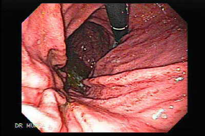 |
Video Endoscopic Sequence 1 of 6.
Endoscopy of Acute Gastritis.
Acute gastritis is not a single disease but, rather, a group of disorders that induce inflammatory changes in the gastric mucosa. All of the causative mechanisms differ in their clinical presentation and have unique histologic characteristics. The inflammation may involve the entire stomach (pangastritis) or a region of the stomach (eg, antral gastritis). Acute gastritis can be broken down into the following additional categories: erosive (eg, hemorrhagic erosions, superficial erosions, deep erosions and nonerosive (generally caused by Helicobacter pylori).

For more endoscopic details download the video clips by clicking on the endoscopic images, wait to be downloaded complete then press Alt and Enter; thus you can observe the video in full screen.
All endoscopic images shown in this Atlas contain
video clips.
|
|
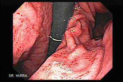 |
Video Endoscopic Sequence 2 of 6.
Endoscopic Picture of Acute Gastritis.
Injury to the gastric mucosa is associated with epithelial cell damage and regeneration. The term gastritis is used to denote inflammation associated with mucosal injury. However, epithelial cell injury and regeneration are not always accompanied by mucosal inflammation. This distinction has caused considerable confusion since gastritis is often used to describe endoscopic or radiologic characteristics of the gastric mucosa rather than specific histologic findings. Epithelial cell damage and regeneration without associated inflammation is properly referred to as "gastropathy".

|
|
 |
Video Endoscopic Sequence 3 of 6.
Endoscopic appearance of Acute Gastritis.
The causes, natural history, and therapeutic implications of gastropathy differ from gastritis:
Gastropathy is usually caused by irritants such as drugs (eg, nonsteroidal antiinflammatory agents and alcohol), bile reflux, hypovolemia, and chronic congestion.
Gastritis is usually due to infectious agents (such as Helicobacter pylori) and autoimmune and hypersensitivity reactions.
Most classification systems distinguish acute, short-term fromchronic, long-term disease. The terms acute and chronic are alsoused to describe the type of inflammatory cell infiltrate. Acuteinflammation is usually associated with neutrophilic infiltration,while chronic inflammation is usually characterized bymononuclear cells, chiefly lymphocytes, plasma cells andmacrophages. A practical clinicopathologic framework for theclassification of gastritis and gastropathy based upon these factorscan be proposed.
|
|
 |
Video Endoscopic Sequence 4 of 6.
Endoscopy of Acute Gastritis.
Acute gastritis may produce no symptoms but can be associated with short-lived dyspepsia, lack of appetite, nausea or vomiting. It can occasionally be severe enough to cause gastrointestinal bleeding with melena or hematemesis (see above). The most common cause is ingestion of aspirin or other non-steroidal anti-imflammatory drugs (NSAIDs). It can also occur during the early stages of infection with the bacteria, Helicobacter pylori "HP." Most cases resolve by themselves, but endoscopy and biopsy may be required to exclude other conditions such as peptic ulcer disease or cancer. At endoscopy the inner lining of the stomach (mucosa) may appear swollen, reddened and inflamed. There may be small, shallow erosions (breaks in the surface lining) or even tiny areas of bleeding from the mucosa.
|
|
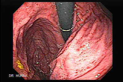 |
Video Endoscopic Sequence 5 of 6.
Acute Gastritis.
Acute gastritis has a number of causes, including certain drugs; alcohol; bacterial, viral, and fungal infections; acute stress (shock); radiation; and direct trauma.
Nonsteroidal anti-inflammatory drugs (NSAIDs), such as aspirin, ibuprofen, and naproxen, can be direct irritants and a cause of gastritis. Because of gravity, the irritants lie on the greater curvature of the stomach, and therefore, gastritis and ulcers are seen distally on or near the greater curvature of the stomach. Gastritis can occur even when NSAIDs are taken systemically or when the drugs are ingested persistently or in large quantities. Alcoholic beverages, such as whisky, vodka, and gin, are direct irritants to the stomach and can cause alcoholic gastritis.
|
|
 |
Video Endoscopic Sequence 6 of 6.
Endoscopy of Acute Gastritis
Bacterial infections also can cause gastritis. The most common type of infection is caused by H pylori. H pylori gastritis typically starts in the antrum, causing intense inflammation, and over time, it may extend to involve the entire gastric mucosa. It is also responsible for as many as 80% of gastric ulcers and is associated with a transient increase in gastric acid secretion. The acid secretion drops as the corpus of the stomach is involved. Urease produced by H pylori catalyzes the hydrolysis of urea to ammonia and carbon dioxide. Hydroxide ions generated by the equilibration of water with ammonia are hypothesized to contribute to gastric mucosal epithelial damage. H pylori is thought to be spread from person to person via oral-oral and/or fecal-oral routes.
|
|
 |
Video Endoscopic Sequence 1 of 10.
Endoscopy of Acute Gastritis (Pangastritis).
The mechanisms of mucosal injury in gastritis and The mechanisms of mucosal injury in gastritis and Peptic ulcer disease are thought to be an imbalance of aggressive factors, such as acid production or pepsin, and defensive factors, such as mucus production, bicarbonate, and blood flow.
|
|

| |
Video Endoscopic Sequence 2 of 10.
Endoscopy of Pangastritis.
H. pylori infection has been associated with a decrease in gastric juice ascorbic acid concentration, and this effect was more pronounced in patients with the CagA-positivestrain. Pangastritis was more common in patients whose H. pylori. infection was accompanied by anemia.
|
|
 |
Video Endoscopic Sequence 3 of 10.
Acute Gastritis.
Gastritis is defined as microscopic inflammation of the stomach and represents a histological not a clinical entity, as the majority of persons with gastric inflammation are completely asymptomatic. A myriad of etiological agents can induce gastritis, but the most common cause is infection by Helicobacter pylori, a bacterium that resides in the mucus gel layer overlying the gastric mucosa.
|
|
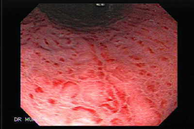 |
Video Endoscopic Sequence 4 of 10.
Video Endoscopy of Acute Gastritis.
The most common causes of acute gastritis are infectious. Acute infection with H. pylori induces gastritis. However, H. pylori acute gastritis has not been extensively studied. Reported as presenting with sudden onset of epigastric pain, nausea, and vomiting, limited mucosal histologic studies demonstrate a marked infiltrate of neutrophils with edema and hyperemia. If not treated, this picture will evolve into one of chronic gastritis. Hypochlorhydria lasting for up to 1 year may follow acute H. pylori infection. The less common of the two forms involves primarily the fundus and body.
|
|
 |
Video Endoscopic Sequence 5 of 10.
Endoscopy of Acute Gastritis.
Acute gastritis may produce no symptoms but can be associated with short-lived dyspepsia, lack of appetite, nausea or vomiting. It can occasionally be severe enough to cause gastrointestinal bleeding with melena or hematemesis.
|
|
 |
Video Endoscopic Sequence 6 of 10.
Acute Gastritis.
In pangastritis (coexisting antral gland gastritis and fundic gland gastritis), the fundic gland gastritis is characterized by less severe inflammation than the antral gland gastritis. Pangastritis can result in extensive mucous gland (pseudopyloric) metaplasia and varying amounts of intestinal metaplasia. Nests of parietal cells commonly occur, even in the presence of achlorhydria.
|
|
 |
Video Endoscopic Sequence 7 of 10.
Endoscopy of Acute Gastritis.
A strong link has been established between H. pylori and a diverse spectrum of gastroduodenal diseases, including gastric and duodenal ulceration, gastric adenocarcinoma, mucosa-associated lymphoid tumor (MALToma), and non-Hodgkin lymphoma of the stomach.
|
|
 |
Video Endoscopic Sequence 8 of 10.
Acute Gastritis.
H. pylori colonizes the stomach for years or decades, not days or weeks, as is usually the case for bacterial pathogens, and this organism virtually always induces chronic gastritis; however, only a fraction of H. pylori -colonized individuals ever develop disease.
|
|
 |
Video Endoscopic Sequence 9 of 10.
Endoscopy of Acute Gastritis.
|
|
 |
Video Endoscopic Sequence 10 of 10.
Endoscopy of Acute Gastritis.
|
|
 |
Severe Acute Gastritis.
Redness of the Mucosa.
Acute inflammation and chronic inflammation usually are distinguishable by general histopathology. In the gastric mucosa, hyperemia and exudation (the endoscopic sign of acute inflammation) The classification of gastritis is based on the intensity of the inflammatory process and its effects on the glandular structures.
|
|
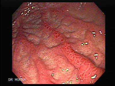 |
Acute Gastritis of the Gastric Fundus.
Reddened Gastric folds.
Fundic (oxyntic) gland gastritis (type A) can be associated with diffuse severe mucosal atrophy and pernicious anemia.
Diffuse severe atrophic fundic gland gastritis primarily affects elderly patients, who develop achlorhydria, but with a residual ability to absorb vitamin B12. Patients with pernicious anemia generally have antibodies against parietal cells (90%) and intrinsic factor (60%). Antiparietal cell antibodies also occur in 50% of patients with gastric atrophy without pernicious anemia (but without anti-intrinsic factor antibodies) and in 10 to 15 % of normal persons.
|
|
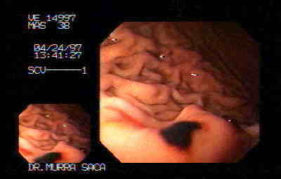 |
Hemorrhagic Gastritis.
Sub epithelial hemorrhages of the stomach are nonspecific entities that may arise in response to a variety of topical insults such as alcohol, corrosives, and drugs (specially non steroidal anti-inflammatory agents) and in response to a severe physiologic stress.
|
|
 |
Hemorrhagic Gastritis.
A 77 year-old, physician with hemorrhagic gastritis of the fundus.
He had been under aspirin treatment.
Acute hemorrhagic gastritis is an important cause of upper gastrointestinal bleeding, accounting for approximately one fourth of UGI bleeding in endoscopic studies. Most patients with hemorrhagic gastritis have underlying predisposing conditions, such as alcohol abuse, portal hypertension, short- or long-term NSAID use, and physiologic stress associated with hospitalization in an ICU for severe life-threatening disease or trauma. The key to management is prevention; however, once established, hemorrhagic gastritis is treated with both supportive measures and measures directed toward healing the mucosal damage. In general, therapy is the same as that for classic peptic ulcer disease. These patients present a challenge, however, because of their underlying diseases and because of the potential for diffuse mucosal bleeding, the latter making the use of endoscopic therapy more difficult. Surgery is an option of last resort for the patient who continues to bleed despite aggressive medical and endoscopic therapy. Future investigations will focus on pharmacologic therapy to enhance mucosal defense mechanisms, therapy that will likely attain increasing importance in the years to come.
|
|
 |
Acute Gatritis of the Antrum.
Antral gland gastritis (type B) is the most common pattern; it is associated with Helicobacter pylori and duodenal ulcers.
In some persons, chronic superficial antral gland gastritis associated with H. pylori progresses to multifocal atrophic gastritis. However, the mechanism by which gastric atrophy occurs is unclear and is probably multifactorial. As the atrophy progresses, the presence of H. pylori decreases, possibly because hypochlorhydria creates an environment that is not conducive to the bacteria.
|
|
 |
Video Endoscopic Sequence 1 of 4.
Endoscopic Picture of Acute Gastritis.
|
|
 |
Video Endoscopic Sequence 2 of 4.
Endoscopic appearance of Acute Gastritis
|
|
 |
Video Endoscopic Sequence 3 of 4.
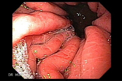
Image and video clip of Severe Acute Gastritis.
|
|
 |
Video Endoscopic Sequence 4 of 4.
Endoscopy of severe acute gastritis.
|
|
 |
Video Endoscopic Sequence 1 of 5.
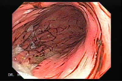
Endoscopy of Subepithelial Hemorrhages
Endoscopy displays severe diffuse subepithelial hemorrhages.
|
|
 |
Video Endoscopic Sequence 2 of 5.
|
|
 |
Video Endoscopic Sequence 3 of 5.
|
|
 |
Video Endoscopic Sequence 4 of 5.
|
|
 |
Video Endoscopic Sequence 5 of 5.
|
|
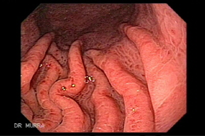 |
Video Endoscopic Sequence 1 of 2.
Image and video clip of Severe Acute Gastritis.
|
|
 |
Video Endoscopic Sequence 2 of 2.
Endoscopy of severe acute gastritis.
|
|
|
|
|
|
|
