 |
|
.
A female Trichuris has been found in the cecum that is the normal habitat of this worms.
Trichuriasis is an intestinal infection found in human beings which is caused by Trichuris trichiura, more commonly known as whipworm because of its whip-like appearance. It is characterized by the invasion of the colonic mucosa by the adult Trichuris and produces minor inflammatory changes at the sites of localization.
It is prevalent throughout the world, especially in tropical areas. Its diagnosis is usually made by identification of the typical eggs in the stool; adult whipworm is rarely seen during colonoscopy. Colonoscopy can directly diagnose trichuriasis, confirming the threadlike form of worms with an attenuated end.
The worms can be overlooked, particularly if colon preparation is imperfect. Attenuated whip-like ends of whipworms, which are embedded in the colonic mucosa, were removed with biopsy forceps.") |
Video Endoscopic Sequence 1 of 19.
Colonoscopic Findings of a Trichuris Trichuris (whipworm).
A female Trichuris has been found in the cecum that is the normal habitat of this worms.
Trichuriasis is an intestinal infection found in human beings which is caused by Trichuris trichiura, more commonly known as whipworm because of its whip-like appearance. It is characterized by the invasion of the colonic mucosa by the adult Trichuris and produces minor inflammatory changes at the sites of localization.
It is prevalent throughout the world, especially in tropical areas. Its diagnosis is usually made by identification of the typical eggs in the stool; adult whipworm is rarely seen during colonoscopy. Colonoscopy can directly diagnose trichuriasis, confirming the threadlike form of worms with an attenuated end.
The worms can be overlooked, particularly if colon preparation is imperfect. Attenuated whip-like ends of whipworms, which are embedded in the colonic mucosa, were removed with biopsy forceps.

Download the video clip by clicking on the endoscopic image. If you wish to observe in full screen, wait to be downloaded complete then press Alt and Enter.
Configure the windows media in repeat is optimal.
All endoscopic images shown in this Atlas contains video clips.
|
|
, which was seen to be moving gently in the cecum, was discovered during the examination.") |
Video Endoscopic Sequence 2 of 19.
This worm seem to be a female, Note: A filariform anterior part. See the histopathologic below.
A whipworm (Trichuris trichiura), which was seen to be moving gently in the cecum, was discovered during the examination.
|
|
 from its characteristic whiplike shape; the adult (male - 30-45 mm, female - 35-50 mm) with males being somewhat smaller than females.
Buries its thin, threadlike anterior half into the intestinal mucosa, After 10-14 days in soil, eggs become infective.
Soil-containing eggs must be swallowed.
No tissue migratory phase occurs. Larvae hatch in the small intestine, where they grow and molt, finally taking up residence in the large intestine.
The time from ingestion of eggs to development of mature worms is approximately 3 months. Adult females lay eggs for up to 5 years.") |
Video Endoscopic Sequence 3 of 19.
Colonoscopic finding of showing a long slender whitish T. trichiura worm in the cecum.
The worm derives its name (whipworm) from its characteristic whiplike shape; the adult (male - 30-45 mm, female - 35-50 mm) with males being somewhat smaller than females.
Buries its thin, threadlike anterior half into the intestinal mucosa, After 10-14 days in soil, eggs become infective.
Soil-containing eggs must be swallowed.
No tissue migratory phase occurs. Larvae hatch in the small intestine, where they grow and molt, finally taking up residence in the large intestine.
The time from ingestion of eggs to development of mature worms is approximately 3 months. Adult females lay eggs for up to 5 years. |
|
 live in the cecum and ascending colon.
The adult worms are fixed in that location with the anterior portions threaded into the mucosa. Most cases are asymptomatic. Heavy parasite loads cause diarrhea, anemia and pain.") |
Video Endoscopic Sequence 4 of 19.
Colonoscopic finding of case 2 showing a whitish worm,
T.trichiura, with coiled posterior end embedded in the wall of the cecum.
Another trichuris is observed in the appendicular hole.
This worm seem to be a male, Note : A filariform anterior part and coiled posterior end.
More than 700 million people are infected worldwide.
The adult worms (approximately 4 cm in length) live in the cecum and ascending colon.
The adult worms are fixed in that location with the anterior portions threaded into the mucosa. Most cases are asymptomatic. Heavy parasite loads cause diarrhea, anemia and pain. |
|
 |
Video Endoscopic Sequence 5 of 19.
Endoscopic parasite extraction and medical treatment was performed.
One of the worm was taken with the biopsy forceps, see below the sequences of video clips and pictures of this whipworm.
Eosinophilia is uncommon; however, when present, it ranges from 5-20%. |
|
 |
Video Endoscopic Sequence 6 of 19.
A video clip and a image of the whipworm.
Macroscopic and histopathologic images are displayed in this sequence, see below. |
|
 |
Video Endoscopic Sequence 7 of 19.
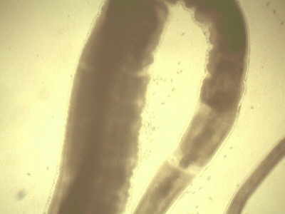
Caudal aspect of female Trichuris trichura.
(Stereoscopic view).
The history of T. Trichuria dates back to the times of prehistoric man; however, the first written record of T.
Trchiura appeared in 1740 when an Italian scientist by the name of Morgani discovered the residence of adult T.
Trichiura worms in the colon. In 1761 Roedere, a German physician, gave a report of the exact morphologic description and provided accurate drawings of the parasite.
The organism received its taxonomy classification in the 18th C. |
|
 |
Video Endoscopic Sequence 8 of 19.
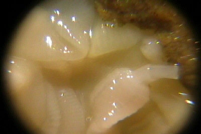
Cephalic part of the Trichuris trichura separated from the body.
To the left there is a portion of caudal part of the parasite.
The mouth is a simple opening, lacking lips.
The buccal cavity is tiny and is provided with a minute spear. |
|
 |
Video Endoscopic Sequence 9 of 19.
Stereoscopic view of the body of the parasite.
The esophagus is very long, occupying about two thirds of the body length and consists of a thin-walled tube surrounded by large, unicellular glands, the stitchocytes. |
|
 |
Video Endoscopic Sequence 10 of 19.
Sections of the worm, demonstrated a gravid female adult nematode with numerous immature ova.
This is a longitudinal section of Trichuris trichura, showing eggs in the uterus.
Female worms in the cecum shed between 3,000-20,000 eggs per day. |
|
 for development of larvae.
When infective eggs are ingested, the larvae hatch in the small intestine, and migrate to the lower intestine and colon, and enters the epithelium of the intestine.
They become sexually mature in about 3 months. The larvae bore into the mucosa with the slender anterior part of the body.") |
Video Endoscopic Sequence 11 of 19.
A complete section of the uterus with numerous eggs.
Life cycle of T.trichiura:
Eggs, after are discharges with the feces, require between 12 days and several weeks (based on the environment condition) for development of larvae.
When infective eggs are ingested, the larvae hatch in the small intestine, and migrate to the lower intestine and colon, and enters the epithelium of the intestine.
They become sexually mature in about 3 months. The larvae bore into the mucosa with the slender anterior part of the body. |
|
 |
Video Endoscopic Sequence 12 of 19.
Characteristic eggs of Trichuris trichura with typical extremes.
Each worm causes an estimated 5 mL of blood loss every day. Heavy infections are required to cause anemia. |
|
 |
Video Endoscopic Sequence 13 of 19.
A female adult T. trichiura, showing the uterine tubules.
A 10x view of eggs into the uterus. |
|
 |
Video Endoscopic Sequence 14 of 19.
A 4x view of eggs.
Whipworm infections transpire as a result of accidental ingestion of Trichuris trichiura eggs or embryos. After they are swallowed, the eggs move to the host's small intestine, where they develop into juveniles.
The young worms then move to the large intestine and attach their anterior ends to the intestinal wall. After approximately three months, the parasite becomes a sexually mature adult, females producing up to 10,000 eggs per day, which are passed out of the host's system with digestive wastes.
The eggs need a warm moist environment to survive outside the body and become infective in about three weeks. Since whipworms do not actually multiply inside a host, each individual worm represents a separate incident of infection. |
|
 |
Video Endoscopic Sequence 15 of 19.
Colonic mucosa with erosion and chronic inflammatory changes, and hemorrhages, 10x. |
|
.") |
Video Endoscopic Sequence 16 of 19.
The colonia mucosae shows chronic inflamation and superficial hemorrage.(Low power 4x). |
|
 |
Video Endoscopic Sequence 17 of 19.
Low power,4x, of colonic mucosa with inflamatory changes.
There are superficial erosions. |
|
 |
Video Endoscopic Sequence 18 of 19.
Low power, 4x, of colonic mucosa with inflamatory changes. |
|
 |
Video Endoscopic Sequence 19 of 19.
Same picture of colonic mucosa at 10x. |
|
 |
Video Endoscopic Sequence 1 of 9.
Schistosoma Mansoni in the Cecum
Magnification Colonoscope.
This 34 year-old male, a routine colonoscopy was performed a Schistosoma Mansoni was found at the cecum.
In the Republic of El Salvador no cases of Schistosoma Mansoni have been reported previously, in this atlas we presented two cases of Schistosoma Mansoni found them at the cecum. |
|
. This Acute Schistosomiasis is not however as important as the chronic forms of the disease. For S. mansoni and S.
Japonicum these are Intestinal and Hepatic Schistosomiasis, associated with formation of granulomas around trapped eggs lodged in the intestinal wall or in the liver respectively.
The hepatic form of the disease is the most important, granulomas here giving rise fibrosis of the liver and hepatosplenomegaly in severe cases.") |
Video Endoscopic Sequence 2 of 9.
High Magnification Image and Video Clip.
The Schistosome Egg is Responsible For Most of the Pathology Associated with Schistosomiasis.
Schistosome eggs, which may become lodged within the hosts tissues, are the major cause of pathology in schistosomiasis, which takes a number of forms.
Onset of egg laying in humans is sometimes associated with an onset of fever (Katayama fever). This "Acute Schistosomiasis" is not however as important as the chronic forms of the disease. For S. mansoni and S.
Japonicum these are "Intestinal" and "Hepatic Schistosomiasis", associated with formation of granulomas around trapped eggs lodged in the intestinal wall or in the liver respectively.
The hepatic form of the disease is the most important, granulomas here giving rise fibrosis of the liver and hepatosplenomegaly in severe cases. |
|
 is a human disease syndrome caused by infection from one of several species of parasitic trematodes of the genus Schistosoma.
Schistosomiasis is a major source of morbidity and mortality for developing countries in Africa, South America, the Caribbean, the Middle East, and Asia.
Tourism to and immigration from endemic areas can result in schistosomiasis cases presenting anywhere in the developed world.") |
Video Endoscopic Sequence 3 of 9.
Schistosomiasis (ie, bilharziasis) is a human disease syndrome caused by infection from one of several species of parasitic trematodes of the genus Schistosoma.
Schistosomiasis is a major source of morbidity and mortality for developing countries in Africa, South America, the Caribbean, the Middle East, and Asia.
Tourism to and immigration from endemic areas can result in schistosomiasis cases presenting anywhere in the developed world. |
|
.") |
Video Endoscopic Sequence 4 of 9.
Most human schistosomiasis is caused by Schistosoma haematobium, Schistosoma mansoni, or Schistosoma japonicum.
Less prevalent species such as Schistosoma mekongi and Schistosoma intercalatum also may cause systemic human disease.
Less importantly, other schistosomes with avian or mammalian primary hosts can cause severe dermatitis in humans (eg, swimmer's itch secondary to Trichobilharzia ocellata). |
|
 reports estimate that 500-600 million people in 74 tropical and subtropical countries are at risk for schistosomiasis.
Over 200 million people in these countries are infected. Of these, 120 million are symptomatic, with 20 million having severe clinical disease.")
Video Endoscopic Sequence 5 of 9.
After malaria, schistosomiasis is the second most prevalent tropical disease in the world. In some parts of the world, it also is known as bilharzia in honor of Theodore Bilharz. He first identified the etiological agent for Schistosoma hematobium in Egypt in 1851.
The pathophysiology of schistosomiasis is due to the immune response against the schistosome eggs. The clinical manifestations depend on the species of parasite, intensity of worm burden, and immunity of the person to the parasite. Recent World Health Organization (WHO) reports estimate that 500-600 million people in 74 tropical and subtropical countries are at risk for schistosomiasis.
Over 200 million people in these countries are infected. Of these, 120 million are symptomatic, with 20 million having severe clinical disease. |
|
 |
Video Endoscopic Sequence 6 of 9.
The life cycle of the flatworms that cause human schistosomiasis involves a sexual stage in the human and an asexual stage in the fresh water snail host.
The adult worms are small, 12-26 mm long and 0.3-0.6 mm wide, and vary with the different species.
S hematobium lives in the venous plexus near the urinary bladder and ureters, S mansoni lives in the inferior mesenteric vein, and S japonicum lives in the superior mesenteric vein of both the large and small intestines. |
|
 |
Video Endoscopic Sequence 7 of 9.
Adult worms mate and lay eggs. The eggs are nonoperculate, possess a spine, and contain a miracidium.
The microscopic appearance of the egg allows diagnostic differentiation of the 5 species.
An adult S hematobium produces 20-200 round, terminally spined eggs per day; S mansoni produces 100-300 ovoid, laterally spined eggs per day; and S japonicum produces 500-3500 round, small, laterally spined eggs per day. The eggs of S intercalatum have prominent terminal spines, and S mekongi have small lateral spines. |
|
 |
Video Endoscopic Sequence 8 of 9.
Pathophysiology: The pathophysiology of infection correlates with the life cycle of the parasite, as follows.
Cercariae: Skin penetration of cercariae produces an allergic dermatitis at the site of entry. With prior sensitization, a pruritic papular rash occurs. This also is observed with nonhuman avian schistosomes.
Schistosomula: These are tailless cercariae that are transported through blood or lymphatics to the right side of the heart and lungs. Heavy infection can cause symptoms such as cough and fever. Eosinophilia may be observed.
Adult worm: Adult worms do not multiply inside the human body. In the venous blood, adult male and female worms mate, and the female lays eggs 4-6 weeks after cercarial penetration. Adult worms rarely are pathogenic.
The female adult worm lives for approximately 3-8 years and lays eggs throughout her life span.
Eggs: They cause Katayama fever and schistosomiasis.
Katayama fever: The exact pathophysiology is not known. It occurs 4-6 weeks after infection, at the time of the initial egg release. It is reported most commonly with S japonicum but also has been reported with S mansoni.
Katayama fever is believed to be due to the high worm and egg antigen stimuli that result from immune complex formation and lead to a serum sickness–like illness. This syndrome is not due to granuloma formation.
Schistosomiasis: It is due to immunological reactions to Schistosoma eggs trapped in tissues. Antigens released from the egg stimulate a granulomatous reaction comprised of T cells, macrophages, and eosinophils that results in clinical disease.
Symptoms and signs depend on the number and location of eggs trapped in the tissues. Initially, the inflammatory reaction is readily reversible. In the latter stages of the disease, the pathology is associated with collagen deposition and fibrosis, resulting in organ damage that may be only partially reversible. |
|
 |
Video Endoscopic Sequence 9 of 9.
The geographic distribution and pathophysiology of schistosomiasis reflect the unique life cycle of these parasites. Schistosomes infect particular species of susceptible freshwater snails in endemic areas.
The infected snails release cercariae, which are fork-tailed free -swimming larvae approximately 1 mm in length. The cercariae survive in freshwater up to 48 hours, during which time they must attach to human skin or to that of another susceptible host mammal or die.
Cercariae attach to human hosts utilizing oral and ventral suckers. They then migrate through intact skin to dermal veins and, over the next several days, to the pulmonary vasculature.
During this migration, the cercariae metamorphose, shedding tails and outer glycocalyces while developing double-lipid-bilayer teguments that are highly resistant to host immune responses. |
|
 |
Video Endoscopic Sequence 1 of 9.
Melanosis Coli and Schistosoma Mansoni
This 63 year-old female a colonoscopy was performed due to an abdominal pain. |
|
 |
Video Endoscopic Sequence 2 of 9.
In spite of to have tried to move the parasite with the pressure of the water spurt, but this action was impossible. |
|
 they begin to mature into miracidia. Eggs that are not shed successfully may remain in the tissues or be swept back to the portal circulation (from the mesenteric vessels) or to the pulmonary circulation (from the vesicular vessels via the inferior vena cava [IVC]).
The free-swimming miracidia that are shed into freshwater survive 2-3 weeks, during which time they must infect a susceptible snail to complete the life cycle. Within the infected snail, 2 generations of sporocysts multiply, mature into free-swimming cercariae, and exit the snail to seek a human host and begin a new cycle.") |
Video Endoscopic Sequence 3 of 9.
The eggs, which are highly antigenic and can induce an intense granulomatous response, migrate through the bowel or bladder wall to be shed via feces or urine.
During this time (approximately 10 d) they begin to mature into miracidia. Eggs that are not shed successfully may remain in the tissues or be swept back to the portal circulation (from the mesenteric vessels) or to the pulmonary circulation (from the vesicular vessels via the inferior vena cava [IVC]).
The free-swimming miracidia that are shed into freshwater survive 2-3 weeks, during which time they must infect a susceptible snail to complete the life cycle. Within the infected snail, 2 generations of sporocysts multiply, mature into free-swimming cercariae, and exit the snail to seek a human host and begin a new cycle. |
|
, and granuloma formation around eggs trapped in the liver and intestinal wall. It resembles 'serum sickness' (i.e. acute immune complex disease), with hepatosplenomegaly, and leucocytosis with eosinophilia.
This phase of the infection is often asymptomatic, but when symptoms do occur they include fever, nausea, headache, an irritating cough and, in extreme cases diarrhoea accompanied with blood, mucus and necrotic material.
These symptoms , if present, last from a few weeks, to several months. It is not as commonly associated with S. mansoni infections compared with those of S. japonicum.") |
Video Endoscopic Sequence 4 of 9.
Acute Schistosomiasis - Also called 'Katayama' fever. This is associated with the onset of the female parasite laying eggs, (approximately 5 weeks after infection), and granuloma formation around eggs trapped in the liver and intestinal wall. It resembles 'serum sickness' (i.e. acute immune complex disease), with hepatosplenomegaly, and leucocytosis with eosinophilia.
This phase of the infection is often asymptomatic, but when symptoms do occur they include fever, nausea, headache, an irritating cough and, in extreme cases diarrhoea accompanied with blood, mucus and necrotic material.
These symptoms , if present, last from a few weeks, to several months. It is not as commonly associated with S. mansoni infections compared with those of S. japonicum. |
|
. The reasons for the high prevalence of this aspect of pathology in Egypt are not clear.") |
Video Endoscopic Sequence 5 of 9.
The Chronic Phase of Infection.
This is the more important aspect of Schistosoma mansoni pathology, and may be divided into two areas Intestinal Schistosomiasis - This and the hepatic schistosomiasis detailed below, manifest a number of years after infection.
The pathogenic reaction is a cellular, granulomatous inflammation around eggs trapped in the tissues, with subsequent fibrosis. All areas of both the small and large intestine may be involved, with the large intestine showing the most severe lesions, whereas severe pathology in the small intestine is only rarely observed, even though large numbers of eggs may be deposited here.
Colonic polyps are also sometimes seen, especially in Egypt (Cheever et al 1978). The reasons for the high prevalence of this aspect of pathology in Egypt are not clear. |
|
 |
Video Endoscopic Sequence 6 of 9.
Hepatosplenic Schistosomiasis - Again, this aspect of the disease is only seen a few years after infection.
The pathology is similar to that seen in the intestine, with a cellular, granulomatous inflammation around eggs trapped in the liver, leading to fibrosis and hepatosplenic disease.
Other organs may more rarely also contain granulomas around eggs, particularly the lungs. |
|
 |
Video Endoscopic Sequence 7 of 9.
Pathology Associated with Infection by Other Species of Schistosome.
S. Japonicum Infection
The primary cause of pathology here is a granulomatous reaction to egg trapped in the liver, and both the acute and chronic aspects of the disease are similar to that of S.
Mansoni infections, although the acute disease Katayama fever, is more common here than for S. mansoni .
The chronic stage of the disease may also be more severe, owing to the greater egg output and longevity of S.
Japonicum females compared with those of S. mansoni. |
|
, hence the infection is often refered to as 'Urinary Schistosomiasis '.
Cancer of the bladder is an important complication of infection with S. haematobium.
Eggs may be deposited in the liver, often in high numbers, and granuloma formation may occur, but this is much less severe than with S. mansoni.") |
Video Endoscopic Sequence 8 of 9.
Schistosoma Mansoni in the cecum
S. haematobium Infection
Adult parasites are found in small venules around the bladder and ureter, with the majority of egg deposition in the tissues of these organs, as eggs pass through the bladder wall, to leave the body in the urine.
The disease cause is chronic in nature, with the most frequently affected organ being the urinary bladder, where calcification of eggs trapped in the tissues often occurs.
The disease is characterised by blood in the urine (haematuria ), hence the infection is often refered to as 'Urinary Schistosomiasis '.
Cancer of the bladder is an important complication of infection with S. haematobium.
Eggs may be deposited in the liver, often in high numbers, and granuloma formation may occur, but this is much less severe than with S. mansoni.
|
|
.
Blood may be seen in the faeces, and diarrhoea may occur. However the portal hypertension . seen in S. mansoni infections has not been reported, and infections are often asymptomatic.
S. mekongi infection.
This has not been greatly studied, but the pathology appears to be similar to that of S. japonicum.") |
Video Endoscopic Sequence 9 of 9.
Minor Species
-
S. Intercalatum infection.
-
This is diagnosed by the presence of terminally spined eggs in the faeces, (the other terminally spined schistosome eggs, those of S. haematobium , are only found in the urine).
Blood may be seen in the faeces, and diarrhoea may occur. However the portal hypertension . seen in S. mansoni infections has not been reported, and infections are often asymptomatic.
-
S. mekongi infection.
-
This has not been greatly studied, but the pathology appears to be similar to that of S. japonicum.
|
|
.
This parasite is prevalent in the tropics, larvae penetrate the intestinal mucosa, and reattach as adults to the mucosa of the cecum wall of the colon. The mucosa becomes edematous and friable. Bleeding from friable mucosa can result in anemia.
This parasite, which only infect human, found in most parts of the world, but mostly in worm and humid areas. The infection is mostly found from children and infection rates as high as 90% has been reported from many areas.") |
Video Endoscopic Sequence 1 of 2.
Trichuris Trichuris (whipworm).
This parasite is prevalent in the tropics, larvae penetrate the intestinal mucosa, and reattach as adults to the mucosa of the cecum wall of the colon. The mucosa becomes edematous and friable. Bleeding from friable mucosa can result in anemia.
This parasite, which only infect human, found in most parts of the world, but mostly in worm and humid areas. The infection is mostly found from children and infection rates as high as 90% has been reported from many areas. |
|
 at the cecum.
Colonoscopy revealed a whitish threadlike worm undulating or coiled in the cecum.
The adult worm usually reaches 3-5 cm in length and has a lifespan of 1-3 years.
Humans are the only known host of T trichiura. The organism is spread via the fecal-oral route. Potential hosts ingest the embryonated (mature) eggs. The eggs hatch in the small intestine, and the larvae attach to and penetrate the small intestinal mucosa, where they begin to mature.
After approximately 1 week, the immature worms move passively to the large intestine and proximal colon. The worms' anterior portions penetrate the mucosal epithelium, and the worms can imbed over one half of their length into the mucosal surface.")
| |
Video Endoscopic Sequence 2 of 2.
Trichuris Trichuris (whip worm) at the cecum.
Colonoscopy revealed a whitish threadlike worm undulating or coiled in the cecum.
The adult worm usually reaches 3-5 cm in length and has a lifespan of 1-3 years.
Humans are the only known host of T trichiura. The organism is spread via the fecal-oral route. Potential hosts ingest the embryonated (mature) eggs. The eggs hatch in the small intestine, and the larvae attach to and penetrate the small intestinal mucosa, where they begin to mature.
After approximately 1 week, the immature worms move passively to the large intestine and proximal colon. The worms' anterior portions penetrate the mucosal epithelium, and the worms can imbed over one half of their length into the mucosal surface. |
|
 may be helpful in these cases.") |
Video Endoscopic Sequence 1 of 8.
Colonoscopy observing a trichuris in the cecum
This 65 year-old female, has been presented during 6 months with diarrheal syndrome, a colonoscopy was performed finding this parasite in the cecum, after specific treatment the patient, never again suffer from diarrhea.
Ingested eggs hatch in the small intestine and complete a 1- to 3-monthmaturation in the large intestine; the cecum and colon are the most commonly infected sites. Adult females lay 5000 to 7000 eggs per day and can do so for up to 5 years.
Diagnosis is by detecting in the feces the oval, yellowish-brown, thick-shelled eggs that have 2 polar plugs. Trichuris eggs can be shed intermittently, so repeated fecal testing or proctoscopy (or colonoscopy, as in the current case) may be helpful in these cases. |
|
 |
Video Endoscopic Sequence 2 of 8.
Colonoscopy of Trichuris Trichuris (whipworm).
Colonoscopy revealed no abnormalities except for a small, white, mobile whip-like worm attached to the cecum. Note the Appendix hole with the whipworm strongly adhered to the mucosa. |
|
 |
Video Endoscopic Sequence 3 of 8.
The whipworm in their lengthwise
The parasite was extracted with biopsy forceps and sent to pathologist for their respective review. |
|
 |
Video Endoscopic Sequence 4 of 8.
The pathologist performed a hematoxylin and eosin staining of the secretion surrounding the parasite, the eggs are microscopically observed that are characteristic of whipworm. |
|
 |
Video Endoscopic Sequence 5 of 8.
Whipworm eggs
A close up to the eggs |
|
 |
Video Endoscopic Sequence 6 of 8.
Multiple eggs are observed |
|
 |
Video Endoscopic Sequence 7 of 8.
Part of the parasite observed under the microscope |
|
 |
Video Endoscopic Sequence 8 of 8.
The whipworm after removal from the cecum |
|
 |
Video Endoscopic Sequence 1 of 6.
Trichuris trichiura Infection Diagnosed by Colonoscopy
This 72 year old male, father of urologist was referred to our endoscopic unit due to discharged a primary tumor of the digestive system because, had been diagnosed with liver metastatic disease and probably a primary kidney tumor, however, no tumor was found in the digestive system but only this whipworm. |
|
 |
Video Endoscopic Sequence 2 of 6.
Another image and video of a whipworm
The adult T. trichiura invade the mucosa and produce minor inflammatory changes at localized sites. In endemic areas, most people are colonized by small numbers of worms and have no symptoms.
Some people harbor hundreds or even thousands of worms, and they present with anemia, diarrhea, abdominal pain, weight loss, malnutrition, appendicitis, colonic obstruction, perforation, or intestinal bleeding. |
|
 |
Video Endoscopic Sequence 3 of 6.
Trichuris trichiura Infection Diagnosed during a screening colonoscopy
Frequency
United States
Whipworm infection is rare overall but is more common in the rural Southeast, where 2.2 million people are thought to be infected.
International
Whipworm infection is more common in less-developed countries. This parasite is carried by nearly one quarter of the world population. |
|
 |
Video Endoscopic Sequence 4 of 6.
Colonoscopic finding of case 3 showing a movable whitish worm, T. trichiura, in the ileocecal valve.
Most patients are asymptomatic. Clinical symptoms are limited to patients with heavy infection, who tend to be small children or others with significant exposure.
Note that there is no pulmonary migration and, thus, no pulmonary or extra-gastrointestinal symptoms.
-
Nocturnal loose stools
-
Dysentery can occur in patients with greater than 200 worms.
-
Rectal prolapse
Failure to thrive
-
Symptoms of anemia (massive infection only)
-
Vague abdominal discomfort
-
Stunted growth
|
|
 |
Video Endoscopic Sequence 5 of 6.
The video shows the pressure exerted by the forceps biopsy removing this parasite which this strongly adhered to the mucosa. |
|
.") |
Video Endoscopic Sequence 6 of 6.
Causes
Whipworm is caused by consumption of soil or food that has been fecally contaminated. (Eggs are infective or embryonated about 2-3 weeks after being deposited in the soil). |
|
 |
Video Endoscopic Sequence 1 of 10.
Trichuris Trichuris (whipworm).
This 57 year-old female underwent a routine colonoscopy finding this parasite in the cecum.
Trichuriasis is an intestinal infection of human beings caused by ingesting embryonated eggs from the environment. Colonized eggs hatch and enter the crypts of the small intestine as larvae
After 1-3 months of maturation, the parasite migrates to the cecum. In the cecum, the parasite matures, mates, and lays eggs. Adult worms are 3-4 cm in length and have thin, tapered anterior regions, and are thus commonly referred to as whipworms. |
|
 |
Video Endoscopic Sequence 2 of 10.
Video Endoscopy of Trichuris Trichuris (whipworm).
Colonoscopic finding, showing a whitish worm, Trichuris trichiura, with coiled posterior end embedded in the wall of the cecum.
Poor hygiene is associated with trichuriasis transmission, and children are especially vulnerable because of their high exposure risk.
This is especially true in developing countries, where poor sanitary conditions correlate with heavy disease burden and infections. |
|
.
In retrospect, the thin head (whip) portion of the worm could be seen embedded in the cecal mucosa while the thicker portion (whip-handle) was visible within the lumen.") |
Video Endoscopic Sequence 3 of 10.
Endoscopy Image of Trichuris Trichuris (whipworm).
Trichuris trichiura, commonly referred to as a whipworm, has a worldwide distribution, particularly among countries with warm, humid climates.
The morphology was that of Trichuris trichiura (whipworm).
In retrospect, the thin head (whip) portion of the worm could be seen embedded in the cecal mucosa while the thicker portion (whip-handle) was visible within the lumen. |
|
 is a combination of symptoms, such as mucoid diarrhea and occasional bleeding.
Rectal prolapse can occur in children with extremely high numbers of T. trichiura worms. Treatment recommendations are as follows: albendazole.
(400 mg once daily for 3 days) or mebendazole (100 mg twice daily for 3 days).") |
Video Endoscopic Sequence 4 of 10.
Removing this parasite which this strongly adhered to the mucosa.
The parasite was removed, and its attenuated whiplike end was gradually extracted from the mucosa by a biopsy forceps and sent for parasitologic examination. The worm was a female.
Trichuris dysentery syndrome (TDS) is a combination of symptoms, such as mucoid diarrhea and occasional bleeding.
Rectal prolapse can occur in children with extremely high numbers of T. trichiura worms. Treatment recommendations are as follows: albendazole.
(400 mg once daily for 3 days) or mebendazole (100 mg twice daily for 3 days). |
|
 |
Video Endoscopic Sequence 5 of 10.
A female adult T. trichiura recovered from the cecum
On a related and fascinating note, Crohn’s disease is uncommon in parts of the world where helminthic colonization is widespread.
Crohn’s disease involves overreactive TH1 pathways and helminths blunt TH1 responses. Because of this and other protective actions, Trichuris suis, the porcine whipworm, has been used successfully to treat Crohn’s disease.
It may not be that the only good helminth is a dead helminth. Sometimes we benefit from sharing a meal with someone who sits next to us. |
|
 |
Video Endoscopic Sequence 6 of 10.
Female adult trichuris trichiura
The worms can be overlooked, particularly if colon preparation is imperfect. |
|
 |
Video Endoscopic Sequence 7 of 10.
Sections of the worm, demonstrated a gravid female adult nematode with numerous immature ova (D, H&E).
Arrel-shaped eggs of T. trichiura, the body wall consisting of the cuticle, epicuticle, and muscle layer (H-E stain). |
|
 |
Video Endoscopic Sequence 8 of 10.
Image of Female Adult Trichuris Trichiura
Almost a fourth of the world’s population is infected withwhipworm, Trichuris trichiura, which, fortunately, is characterized by a relative lack of symptoms; only patients with heavy infections become symptomatic.
Trichuris eggs are unembryonated and not infectious when they are excreted. Development to the infectious stage takes at least 2 weeks, and the host only becomes infected after ingesting embryonated eggs from the environment, usually from dirt or possibly contaminated water. |
|
 |
Video Endoscopic Sequence 9 of 10.
Moderate eosinophilic infiltration in lamina propria |
|
 |
Video Endoscopic Sequence 10 of 10.
Moderate eosinophilic infiltration in lamina propria |
|
 |
Another tiny parasite attached to the cecum
This morphology is different from those presented here in the others sequences, is smaller and brown.
The parasite may be a variant or another whipworm if you have any ideas please send us an email.
|
|
 |
Video Endoscopic Sequence 1 of 3.
Colonoscopy in which there is a whipworm in the sigmoid.
This is a 80 year-old, male. A colonoscopy was practiced because of changes in colonic motility, constipation, insomuch that defecated without laxatives, could spend up to 20 days without a bowel movement. |
|
 |
Video Endoscopic Sequence 2 of 3.
In these videos you can watch this whipworm the suction exerted on the colonic mucosa.
|
|
.
Are observed mucosal lesions apparently caused by the parasite sucking on the mucosa.") |
Video Endoscopic Sequence 3 of 3.
Video Endoscopy of Trichuris Trichuris (whipworm).
Are observed mucosal lesions apparently caused by the parasite sucking on the mucosa. |
|
 |
Video Endoscopic Sequence 1 of 6.
Trichuriasis; Trichuris.
This is the case of a lady of 50 year-old, in a routine colonoscopy a whipworm was found, looking for more parasites colonoscope is advanced to the terminal ileum, but can not find more. only one of this video and image.
laxatives usually expel parasites for this reason the colonoscopists rarely found.
|
|
 |
Video Endoscopic Sequence 2 of 6.
| Colonoscopy of a whipworm |
|
|
|
|
|
.") |
Video Endoscopic Sequence 3 of 6.
Colonoscopy of Trichuris Trichuris (whipworm).
|
|
.") |
Video Endoscopic Sequence 4 of 6.
Colonoscopic Findings of a Trichuris Trichuris (whipworm).
|
|
.") |
Video Endoscopic Sequence 5 of 6.
Colonoscopic Findings of a Trichuris Trichuris (whipworm). |
|
.") |
Video Endoscopic Sequence 6 of 6.
Colonoscopy of Trichuris Trichuris (whipworm).
|
|
 |
Video Endoscopic Sequence 1 of 2.
Colonoscopic Findings of a Trichuris Trichuris in the cecum and melanosis coli
This a 54 year-old, male who in a routine colonoscopy is found this parasite in the cecum, melanosis coli signs are observed, had been consuming Over-the-counter laxatives.
|
|
.") |
Video Endoscopic Sequence 2 of 2.
Melanosis Coli and Trichuris Trichuris (whipworm).
|
|
. Usually asymptomatic, ascariasis is most prevalent in children of tropical and developing countries, where they are perpetuated by contamination of soil by human feces or use of untreated feces as fertilizer.
The parasite is acquired through ingestion of embryonated eggs. Ascariasis is usually asymptomatic but can be complicated by several conditions, including appendicitis, bowel perforation, cholecystitis, intestinal obstruction (large numbers), malabsorption (eg, lactose, nitrogen, vitamin A), and pancreatitis. The mortality rate is 5% if complications occur. When the parasite migrates through the lung early in its parasitic cycle, it can also cause pneumonitis.") |
Video Endoscopic Sequence 1 of 2.
Ascaris lumbricoides
Ascariasis is the most common helminthic infection, with an estimated worldwide prevalence of 25% (0.8-1.22 billion people). Usually asymptomatic, ascariasis is most prevalent in children of tropical and developing countries, where they are perpetuated by contamination of soil by human feces or use of untreated feces as fertilizer.
The parasite is acquired through ingestion of embryonated eggs. Ascariasis is usually asymptomatic but can be complicated by several conditions, including appendicitis, bowel perforation, cholecystitis, intestinal obstruction (large numbers), malabsorption (eg, lactose, nitrogen, vitamin A), and pancreatitis. The mortality rate is 5% if complications occur. When the parasite migrates through the lung early in its parasitic cycle, it can also cause pneumonitis.
People infected with Ascaris often show no symptoms. If symptoms do occur they can be light and include abdominal discomfort. Heavy infections can cause intestinal blockage and impair growth in children. Other symptoms such as cough are due to migration of the worms through the body. Ascariasis is treatable with medication prescribed by your health care provider.
Ascaris lives in the intestine and Ascaris eggs are passed in the feces of infected persons. If the infected person defecates outside (near bushes, in a garden, or field), or if the feces of an infected person are used as fertilizer, then eggs are deposited on the soil. They can then mature into a form that is infective. Ascariasis is caused by ingesting infective eggs. This can happen when hands or fingers that have contaminated dirt on them are put in the mouth or by consuming vegetables or fruits that have not been carefully cooked, washed or peeled.
Adult worms move throughout the GI tract and move in and out of orifices (eg, biliary tract, pancreas, appendix, diverticula, Meckel diverticulum) and may become incarcerated, leading to obstructive pathology. The worms may die, leading to inflammation, necrosis, infection, and abscess formation. If they migrate through an existing perforation in the bowel wall secondary to tuberculosis or typhoid, they can cause a granulomatous peritonitis. Larvae during migration may be deposited in the brain, spinal cord, kidney, or other organs, leading to granuloma formation, inflammation, or infection. They may become entwined in a bolus and obstruct the small bowel; this is most common in the terminal ileum, although other, more proximal, sites have been rarely reported.
|
|
; in fatal cases, the average worm burden was 659.
Based on numerous reports in the literature, a rough estimate of the occurrence (percent of total complications) of complications is as follow") |
Video Endoscopic Sequence 2 of 2.
Ascaris lumbricoides
Female and Male (the smallest)
|
Morbidity is proportional to the worm burden. A large majority of cases are asymptomatic. Intestinal obstruction, the most common complication of ascariasis, has been reported with as few as 4 worms. The average worm burden in numerous nonfatal case reports was 59 worms (range, 4-990); in fatal cases, the average worm burden was 659.
Based on numerous reports in the literature, a rough estimate of the occurrence (percent of total complications) of complications is as follows:[
-
Intestinal obstruction - 63%
-
Bile duct obstruction - 23%
-
Perforation, peritonitis, or both - 3.2%
-
Volvulus - 2.7%
-
Hepatitic abscess - 2.1%
-
Appendicitis - 2.1%
-
Pancreatitis - 1%
-
Cerebral encephalitis - 1%
-
Intussusception - 0.5%
Other sites of pathology (< 0.5%) include Meckel diverticulum, the gallbladder, ears, eyes, nose, lungs, kidneys, vagina, urethra, heart, placenta, spleen, thoracic cavity, umbilicus, pericecal mass, jejunostomy tube, and erythema nodosum. In endemic regions, ascariasis is a significant part of the differential diagnosis for intestinal obstruction, appendicitis, biliary tract disease, pancreatitis, intussusception, and volvulus.
|
|
|
|
 |
Video Endoscopic Sequence 1 of 3.
Taenia Solium
Taenia solium is the pork tapeworm belonging to cyclophyllid cestodes in the family Taeniidae. It is an intestinal zoonotic parasite found throughout the world, and is most prevalent in countries where pork is eaten. The adult worm is found in humans and has a flat, ribbon-like body, which is white in color and measures 2 to 3 m in length. Its distinct head, the scolex, contains suckers and a rostellum as organs of attachment. The main body, the strobila, consists of a chain of segments known as proglottids. Each proglottid is a complete reproductive unit; hence, the tapeworm is a hermaphrodite. It completes its life cycle in humans as the definitive host and pigs as intermediate host. It is transmitted to pigs through human faeces or contaminated fodder, and to humans through uncooked or undercooked pork. Pigs ingest embryonated eggs, morula, which develop into larvae, the oncospheres, and ultimately into infective larvae, cysticerci. A cysticercus grows into an adult worm in human small intestines. Infection is generally harmless and asymptomatic. However, accidental infection in humans by the larval stage causescysticercosis. The most severe form is neurocysticercosis, which affects the brain and is a major cause of epilepsy.
People do not get cysticercosis by eating undercooked pork. Eating undercooked pork can result in intestinal tapeworm if the pork contains larval cysts. Pigs become infected by eating tapeworm eggs in the feces of a human infected with a tapeworm.
|
|
 The severity of cysticercosis depends on location, size and number of parasite larvae in tissues, as well as the host immune response. Other symptoms include sensory deficits, involuntary movements, and brain system dysfunction. In children, ocular location of cysts is more common than cystation in other locations of the body.") |
Video Endoscopic Sequence 2 of 3.
Taenia Solium
Adult T. solium is a , having no body cavity. It is normally 2 to 3 m in length, but can become much larger, sometimes over 8 m long. It is white in colour and flattened into a ribbon-like body. The anterior end is a knob-like head called a scolex, which is 1 mm in diameter.
Intestinal infection of T. solium is called taeniasis which is quite asymptomatic. Only in severe cases, conditions of intestinal irritation, anaemia, and indigestion occur, which can lead to loss of appetite and emaciation. Cysticercus is clinically pathogenic. Ingestion of T. solium eggs or proglottids which rupture within the host intestines can cause larvae to migrate into host tissue to cause cysticercosis. This is the most frequent and severe disease caused by T. solium. In symptomatic cases, a wide spectrum of symptoms may be expressed, including headaches, dizziness, and occasional seizures. In more severe cases, dementia or hypertension can occur due to perturbation of the normal circulation of cerebrospinal fluid. (Any increase in intracranial pressure will result in a corresponding increase in arterial blood pressure, as the body seeks to maintain circulation to the brain.) The severity of cysticercosis depends on location, size and number of parasite larvae in tissues, as well as the host immune response. Other symptoms include sensory deficits, involuntary movements, and brain system dysfunction. In children, ocular location of cysts is more common than cystation in other locations of the body.
In many cases, cysticercosis in the brain can lead to epilepsy, seizures, lesions in the brain, blindness, tumor-like growths, and low eosinophil levels. It is the cause of major neurological problems, such as hydrocephalus, paraplegy, meningitis, convulsions, and even death
|
|
 |
Video Endoscopic Sequence 3 of 3.
Taenia Solium |
|
 Enterobius vermicularis (previously Oxyuris vermicularis) also called human pinworm. (Adult females: 8 to 13 mm, adult male: 2 to 5 mm. ) Humans are considered to be the only hosts of E. vermicularis. A second species, Enterobius gregorii, has been described and reported from Europe, Africa, and Asia. For all practical purposes, the morphology, life cycle, clinical presentation, and treatment of E. gregorii is identical to E. vermicularis.
Eggs are deposited on perianal folds. Self-infection occurs by transferring infective eggs to the mouth with hands that have scratched the perianal area. Person-to-person transmission can also occur through handling of contaminated clothes or bed linens. Enterobiasis may also be acquired through surfaces in the environment that are contaminated with pinworm eggs (e.g. , curtains, carpeting). Some small number of eggs may become airborne and inhaled. These would be swallowed and follow the same development as ingested eggs. Following ingestion of infective eggs, the larvae hatch in the small intestine and the adults establish themselves in the colon. The time interval from ingestion of infective eggs to oviposition by the adult females is about one month. The life span of the adults is about two months. Gravid females migrate nocturnally outside the anus and oviposit while crawling on the skin of the perianal area . The larvae contained inside the eggs develop (the eggs become infective) in 4 to 6 hours under optimal conditions The number 1. Retroinfection, or the migration of newly hatched larvae from the anal skin back into the rectum, may occur but the frequency with which this happens is unknown") |
Video Endoscopic Sequence 1 of 1.
Enterobiasis (also known as Pinworm Infection)
The nematode (roundworm) Enterobius vermicularis (previously Oxyuris vermicularis) also called human pinworm. (Adult females: 8 to 13 mm, adult male: 2 to 5 mm. ) Humans are considered to be the only hosts of E. vermicularis. A second species, Enterobius gregorii, has been described and reported from Europe, Africa, and Asia. For all practical purposes, the morphology, life cycle, clinical presentation, and treatment of E. gregorii is identical to E. vermicularis.
Eggs are deposited on perianal folds. Self-infection occurs by transferring infective eggs to the mouth with hands that have scratched the perianal area. Person-to-person transmission can also occur through handling of contaminated clothes or bed linens. Enterobiasis may also be acquired through surfaces in the environment that are contaminated with pinworm eggs (e.g. , curtains, carpeting). Some small number of eggs may become airborne and inhaled. These would be swallowed and follow the same development as ingested eggs. Following ingestion of infective eggs, the larvae hatch in the small intestine and the adults establish themselves in the colon. The time interval from ingestion of infective eggs to oviposition by the adult females is about one month. The life span of the adults is about two months. Gravid females migrate nocturnally outside the anus and oviposit while crawling on the skin of the perianal area . The larvae contained inside the eggs develop (the eggs become infective) in 4 to 6 hours under optimal conditions The number 1. Retroinfection, or the migration of newly hatched larvae from the anal skin back into the rectum, may occur but the frequency with which this happens is unknown.
|
|
|
|
|
|
|

