 |
|
 |
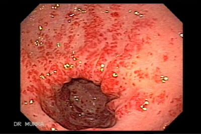
Video Endoscopic Sequence 1 of 9.
Endoscopic appearance of Watermelon Stomach.
70 year-old female, who had history of having anemia and several times with positive fecal-occult-blood had several endoscopy in other clinics, not making the correct diagnosis. Hemoglobin in two years had been fluctuating between 7.0 W / dl to 9.0 W / dl.
The pathogenesis of GAVE syndrome, principally considered an idiopathic disease, is unknown, and theories about it are controversial.
Gastric antral vascular ectasia (GAVE) syndrome, also known as watermelon stomach is a significant cause of acute or chronic gastrointestinal blood loss in the elderly. is characterized endoscopically by “watermelon stripes.” Without cirrhosis, patients are 71% female, average age 73, presenting with occult blood loss leading to transfusion-dependent chronic iron-deficiency anemia, severe acute upper gastrointestinal bleeding, and nondescript abdominal pain.
Download the video clips by clicking on the endoscopic
images, if you wish to observe in full screen, wait to be
downloaded complete then press Alt and Enter for
Windows media, Real Player Ctrl and 3.
Configure the windows media in repeat is optimal.
All endoscopic images shown in this Atlas contain
video clips. We recommend seeing the video clips in full
screen mode.
|
|
 |
Video Endoscopic Sequence 2 of 9.
Video Clip of Watermelon Stomach
Gastric antral vascular ectasia (GAVE) syndrome, also known as watermelon stomach, is a rare but significant cause of severe acute or chronic gastrointestinal blood loss in the elderly. Although it is associated with heterogeneous medical conditions, including hepatic, renal, and cardiac diseases, its pathogenesis is unknown. The diagnosis of GAVE syndrome in patients with renal or hepatic disease is often problematic because there are more frequent causes of gastrointestinal bleeding in these diseases (vascular malformations, peptic ulcer disease, esophageal or gastric varices, and colonic and rectal ulcers) that overshadow GAVE syndrome. Furthermore, diagnosis may be challenging because gastrointestinal bleeding may be occult or overt, and the endoscopic appearance of GAVE syndrome resembles that in portal hypertensive gastropathy (PHG) or antral gastritis. However, differentiation of GAVE syndrome from these other causes is critical because of the vastly disparate therapies required for each. Building on a leading theory that mechanical stress is involved in the pathogenesis, we speculate that the diverse medical risk factors may be related to GAVE syndrome through an autonomic dysfunction.
|
|
 |
Video Endoscopic Sequence 3 of 9.
Gastric antral vascular ectasia (GAVE) syndrome
Endoluminal therapies are the mainstay of conservative management and include endoscopic band ligation, sclerotherapy, heater probe, and argon plasma coagulation, which is emerging as the preferred endoscopic therapy. Although multiple intraluminal treatment sessions may be required for cessation of transfusion dependence, the safety of endoscopic therapy is well documented, and there is only a single case report of a complication —gastric outlet obstruction secondary to argon plasma coagulation. Further, a recent case report described successful endoscopic mucosal resection of lesions in GAVE syndrome with resolution of anemia
Although GAVE syndrome is a rare medical condition, it is a relevant possibility in older patients with severe acute or chronic gastrointestinal blood loss, because it accounts for up to 4% of nonvariceal upper gastrointestinal blood loss. The initial presentation may include occult blood loss leading to transfusion-dependent chronic iron-deficiency anemia, severe acute upper-gastrointestinal bleeding, nondescript abdominal pain, or even gastric outlet obstruction, as described in a prior case report. This disease entity was first described by Rider et al in a patient with severe chronic iron-deficiency anemia and gastroscopy showing “fiery red changes with marked hypertrophic mucosal changes, and scattered profuse bleeding.”
|
|
 |
Video Endoscopic Sequence 4 of 9.
Mallory Weiss Endoscópico
A majority of patients without cirrhosis but with GAVE syndrome are female (71%) with median age of 73 years, whereas the majority of patients with both cirrhosis and GAVE syndrome are male (75%) with a mean age of 65 years. Associated medical conditions include heart, liver, and kidney diseases; diabetes; connective-tissue diseases; hypothyroidism; and status as a bone marrow transplant recipient. The epide-miologic features of GAVE syndrome are attributed to the age and sex distributions of the underlying medical conditions, of which connective-tissue diseases and cirrhosis are the most commonly related. |
|
 |
Video Endoscopic Sequence 5 of 9.
Otra imagen y video del Mallory Weiss Endoscópico.
The syndrome has the name watermelon stomach because of the pathognomonic endoscopic appearance (columns of red tortuous ectatic vessels along longitudinal folds of the antrum) that resembles watermelon stripes Typical histologic changes include superficial hyperplastic antral mucosa, capillary ectasia with thrombosis, and fibromuscular hypertrophy of the lamina propria. GAVE syndrome is often misdiagnosed on endoscopy as PHG. Unlike watermelon stomach, PHG causes predominant changes in the fundus and corpus. GAVE syndrome does not respond to measures that decrease portal pressures in PHG, including transjugular intrahepatic shunt and β-blocker therapy. |
|
 |
Video Endoscopic Sequence 6 of 9.
Hemostatic therapy is initiated with argon plasma coagulation
|
|
 |
Video Endoscopic Sequence 7 of 9.
Argon Plasma in action.
|
|
 |
Video Endoscopic Sequence 8 of 9.
Final view of the treatment with APC
|
|
 |
Video Endoscopic Sequence 9 of 9.
Final view of the treatment with APC
|
|
 |
Video Endoscopic Sequence 1 of 7.
Watermelon Stomach
Longitudinal erythymatous stripes that formed lines within the antrum radiating towards the pylorus resembling the stripes of a watermelon and hence the name gastric antral vascular ectasias (GAVE), or watermelon stomach.
Painless occult gastrointestinal bleeding with anemia in an elderly woman is the most typical presentation. This lesion is amenable to endoscopic thermal ablation, and the lesion shown was treated by argon plasma coagulation.
|
|
 |
Video Endoscopic Sequence 2 of 7.
Treatment of watermelon stomach (GAVE syndrome) with endoscopic argon plasma coagulation (APC).
The diagnosis is based on the endoscopic findings. The typical lesions have longitudinal rugal folds traversing the antrum and converging on the pylorus, each containing a visible convoluted column of vessels, the aggregate resembling the stripes of a watermelon. Although these lesions are confined to the antrum in the majority of cases, up to 33% of the patients have proximal gastric involvement typically in the presence of a diaphragmatic hernia. It is important to emphasize, however, that these lesions might be misdiagnosed as gastritis or portal gastropathy and thus delay in treatment could result.
|
|
 |
Video Endoscopic Sequence 3 of 7.
Watermelon stomach is an increasingly recognizable cause of persistent acute or occult gastrointestinal bleeding, especially in elderly women. The chief presentation is severe iron deficiency anemia and occult or overt gastrointestinal bleeding. Diagnosis is made on endoscopy by the characteristic appearance of visible watermelon linear stripes in the antrum. Histology is rarely needed to confirm the diagnosis. The importance of this lesion lies in the proper recognition since it is amenable to successful therapeutic interventions, leading to endoscopic healing of the lesion, significant improvement in the anemia and a reduction in the need for blood transfusions.
|
|
 |
Video Endoscopic Sequence 4 of 7.
The argon-plasma-coagulation uses instead of laser energy conduction of electric energy by ionized argon gas (plasma), which produces coagulation necrosis of tissues. The potential advantages of the argon-plasma-coagulation lie in the limited deep penetration, which reduces the risk of perforation and the symmetric spread of the coagulation effects in the surrounding mucosa. These properties make the argon plasma-coagulation a promising tool for the endoscopic therapy of mucosal lesions of the GI-tract. Further attractive is the low cost of the argon-plasma -coagulation equipment compared with laser devices.
|
|
 |
Video Endoscopic Sequence 5 of 7.
The therapeutic options are numerous for this condition and needs to be individualized. The simplest form of therapy is iron supplementation and occasional blood transfusions. When these measures fail, other approaches are warranted, including endoscopic, pharmacologic or surgical therapies.
|
|
 |
Video Endoscopic Sequence 6 of 7.
Argon Plasma Coagulator is a new device that allows for non-contact monopolar coagulation of bleeding surfaces, and devitalization of tissue in the gastrointestinal tract. It is safer and much less expensive than lasers, more effective than bipolar cauterization techniques.
|
|
 |
Video Endoscopic Sequence 7 of 7.
The electrode in the argon channel of the probe is connected to an electrosurgical generator.
The APC probe ionizes the argon gas where it remains ionized approximately 2-10mm distal to the tip of the probe. Ionized Argon gas is electrically conductive. This allows the current to flow between the probe and the tissue. Current density upon arrival at the tissue surface causes coagulation. The application of the energy to the tissue is uniform, and contact free. The Argon plasma beam acts not only in a straight line (axially) along the axis of the probe, but also laterally and radially and "around the corner" as it seeks conductive bleeding surfaces. Following physical principles, the plasma beam has a tendency to turn away from already coagulated (high impedance) areas toward bleeding or still inadequately coagulated receiving treatment. This automatically results in evenly applied, uniform surface coagulation.
|
|
 |
Watermelon Stomach
Watermelon-stomach is a rare cause of gastrointestinal bleeding. There has been an increasing number of reports on the association of this lesion with diseases of the scleroderma group.
Gastric antral vascular ectasia (GAVE), also referred to as Watermelon stomach, is a severe haemorrhagic condition that leads to significant morbidity and transfusion dependence in some patients. Re-bleeding following treatment is common, and there are few treatment options. Until recent treatment modalities were developed, the only options available to patients were blood transfusions or the surgical removal of the stomach (antrectomy). The estimated prevalence of GAVE ranges from 0.3 per cent of cases in a large endoscopic series to 4 per cent in highly selected cohorts with severe gastrointestinal bleeding. Although some patients with diffuse GAVE may have portal hypertensive gastropathy, for the purpose of this application the indication is GAVE not related to portal hypertensive gastropathy.
|
|
 |
Video Endoscopic Sequence 1 of 7.
Watermelon Stomach
Longitudinal erythymatous stripes that formed lines within
the antrum radiating towards the pylorus resembling the
stripes of a watermelon and hence the name gastric antral
vascular ectasias (GAVE), or watermelon stomach.
Painless occult gastrointestinal bleeding with anemia in an
elderly woman is the most typical presentation. This lesion
is amenable to endoscopic thermal ablation, and the lesion
shown was treated by argon plasma coagulation.
|
|
 |
Video Endoscopic Sequence 2 of 7.
Treatment of watermelon stomach (GAVE syndrome) with
endoscopic argon plasma coagulation (APC).
The diagnosis is based on the endoscopic findings. The
typical lesions have longitudinal rugal folds traversing the
antrum and converging on the pylorus, each containing a
visible convoluted column of vessels, the aggregate
resembling the stripes of a watermelon. Although these
lesions are confined to the antrum in the majority of cases,
up to 33% of the patients have proximal gastric
involvement typically in the presence of a diaphragmatic
hernia. It is important to emphasize, however, that these
lesions might be misdiagnosed as gastritis or portal
gastropathy and thus delay in treatment could result.
|
|
 |
Video Endoscopic Sequence 3 of 7.
Watermelon stomach is an increasingly recognizable cause of persistent acute or occult gastrointestinal bleeding, especially in elderly women. The chief presentation is severe iron deficiency anemia and occult or overt gastrointestinal bleeding. Diagnosis is made on endoscopy by the characteristic appearance of visible watermelon linear stripes in the antrum. Histology is rarely needed to confirm the diagnosis. The importance of this lesion lies in the proper recognition since it is amenable to successful therapeutic interventions, leading to endoscopic healing of the lesion, significant improvement in the anemia and a reduction in the need for blood transfusions.
|
|
 |
Video Endoscopic Sequence 4 of 7.
The argon-plasma-coagulation uses instead of laser energy
conduction of electric energy by ionized argon gas (plasma),
which produces coagulation necrosis of tissues. The
potential advantages of the argon-plasma-coagulation lie in
the limited deep penetration, which reduces the risk of
perforation and the symmetric spread of the coagulation
effects in the surrounding mucosa. These properties make
the argon plasma-coagulation a promising tool for the
endoscopic therapy of mucosal lesions of the GI-tract.
Further attractive is the low cost of the argon-plasma
-coagulation equipment compared with laser devices.
|
|
 |
Video Endoscopic Sequence 5 of 7.
The therapeutic options are numerous for this condition and
needs to be individualized. The simplest form of therapy is
iron supplementation and occasional blood transfusions.
When these measures fail, other approaches are warranted,
including endoscopic, pharmacologic or surgical therapies.
|
|
 |
Video Endoscopic Sequence 6 of 7.
Argon Plasma Coagulator is a new device that allows for
non-contact monopolar coagulation of bleeding surfaces,
and devitalization of tissue in the gastrointestinal tract. It is
safer and much less expensive than lasers, more effective
than bipolar cauterization techniques.
|
|
 |
Video Endoscopic Sequence 7 of 7.
The electrode in the argon channel of the probe is
connected to an electrosurgical generator.
The APC probe ionizes the argon gas where it remains
ionized approximately 2-10mm distal to the tip of the probe.
Ionized Argon gas is electrically conductive. This allows the
current to flow between the probe and the tissue. Current
density upon arrival at the tissue surface causes
coagulation. The application of the energy to the tissue is
uniform, and contact free. The Argon plasma beam acts not
only in a straight line (axially) along the axis of the probe,
but also laterally and radially and "around the corner" as it
seeks conductive bleeding surfaces. Following physical
principles, the plasma beam has a tendency to turn away
from already coagulated (high impedance) areas toward
bleeding or still inadequately coagulated receiving
treatment. This automatically results in evenly applied,
uniform surface coagulation.
|
|
 |
Video Endoscopic Sequence 1 of 14.
Endoscopy of Case of Stomach in Watermelon and Multiple angiodysplasias of the fundus and duodenum.
Gastric Antral Vascular Ectasias,
A 53-year-old female, with hemoglobin of 7.3 g / dl with liver cirrhosis and hepatocarcinoma. |
|
 |
Video Endoscopic Sequence 2 of 14.
The patient has several angiodysplasias of the gastric fundus
|
|
 |
Video Endoscopic Sequence 3 of 14.
In the duodenum there are some angiodysplasias, which carried out ablative therapy with argon plasma coagulator APC . |
|
 |
Video Endoscopic Sequence 4 of 14.
"Watermelon Stomach" Gastric Antral Vascular Ectasia.
|
|
 |
Video Endoscopic Sequence 5 of 14.
Gastric angiodysplasias
Ablative therapy with argon plasma coagulator is applied
|
|
 |
Video Endoscopic Sequence 6 of 14.
Image and video of Gastric angiodysplasias
|
|
 |
Video Endoscopic Sequence 7 of 14.
Argon plasma coagulation (APC) is an endoscopic procedure used with a high-frequency electrical current for control of bleeding from gastrointestinal vascular ectasias including angiodysplasia and gastric antral vascular ectasia.
Image and video of ablative therapy with argon plasma coagulator.
|
|
 |
Video Endoscopic Sequence 8 of 14.
Image and video of ablative therapy with argon plasma coagulator.
|
|
 |
Video Endoscopic Sequence 9 of 14.
Final Status of coagulation with argon plasma in watermelon stomach.
|
|
 |
Video Endoscopic Sequence 10 of 14.
Final Status of coagulation with argon plasma in watermelon stomach.
|
|
 |
Video Endoscopic Sequence 11 of 14.
One week later (2nd session)
It Repeated ablative therapy with argon plasma coagulator
The improvement is remarkable have been lessened vascular lesions with the first treatment. |
|
 |
Video Endoscopic Sequence 12 of 14.
Endoscopic Therapeutics with APC
|
|
 |
Video Endoscopic Sequence 13 of 14.
Treatment with APC (2nd session). |
|
 |
Video Endoscopic Sequence 14 of 14.
Patient with liver cirrhosis and hepatocellular carcinoma.
Final state of coagulation with argon plasma in stomach in watermelon (second treatment). |
|
Estomago en Sandia |
|
|
|
|
