 |
|
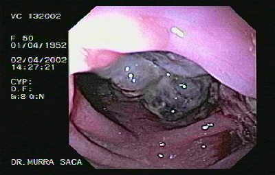 |
Video Endoscopic Sequence 1 of 11.
Metastasis of Renal Carcinoma to the ascending colon. Multilobulated Neoplasia of the ascending colon near the cecum which manifested as a primary colonic tumour.
A 50 year-old female with rectal bleeding and weight loss of more than 20 pounds. 4 months previously patient had a negative barium enema. The patient underwent a unilateral nephrectomy, 3 years previously due to renal carcinoma of the right kidney.
Colon metastasis of renal cell carcinoma is very rare.
We recently had a case of Gastric Metastasis from Renal Cell Carcinoma, And morphologically resembles the case presented hereto see this case Click here
For more endoscopic details download the video clip by clicking on the endoscopic image. Wait to be downloaded complete, then if you wish to appreciate in full screen press Alt and Enter.
All endoscopic images of this atlas contain a video clip.
|
|
 |
Video Endoscopic Sequence 2 of 11.
Solitary colonic metastasis from renal cell carcinoma 3 years after nephrectomy.
Right hemicolectomy was performed. Postoperative histological examination revealed that the tumour was a metastatic renal cell carcinoma of the clear cell type.
A large encapsulated mass is observed.
The cecum was explored. The video clip also displays the orifice of appendix.
|
|
 |
Video Endoscopic Sequence 3 of 11.
A large encapsulated mass is observed.Metastases of Renal Carcinoma to Ascending Colon. When the biopsy was taken an encapsulated tumor was observed. The tumor´s morphology was of a membrane-like capsule, which lead to the impression that the mass was not a typical carcinoma but of a different etiology.
|
|
 towards the lower border of the liver, without infiltrating that organ.") |
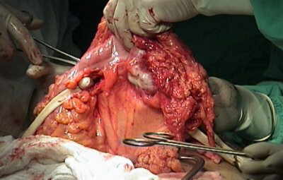
Video Endoscopic Sequence 4 of 11.
The multilobulated tumor is observed. Bleeding is appreciated. During surgery, an invasive neoplastic tumor was found which extended from a displaced cecum, (due to prior nephrectomy) towards the lower border of the liver, without infiltrating that organ.
|
|
 |
Video Endoscopic Sequence 5 of 11.
Bleeding is observed due to the fragility of the tumor.
Renal cell carcinoma is the most common primary neoplasm of the kidney. Concomitant metastasis of renal cell carcinoma is seen in 30% of patients at the time of diagnosis. Those are mostly seen in the lungs, lymph nodes, liver and brain. Late metastasis can develop years after the curative nephrectomy in 50% of patients but colonic metastasis is very rare.
|
|
.") |
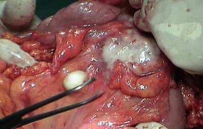
Video Endoscopic Sequence 6 of 11.
This picture displays the surgical specimen of that metastases at the operation room. (whitish part).
|
|
 |
Sequence 7 of 11.
An ileo-transverse anastomosis was performed.
The Pre and postoperative histologic examination revealed
that the tumor was a metastatic clear cell carcinoma.
|
|
 |
Sequence 8 of 11.
Metastases of Carcinoma Renal to the ascending colon
A close up to the neoplasia.
|
|
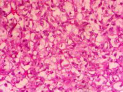 |
Sequence 9 of 11.
A high power magnification shows the vacuolated clear
cell cytoplasm of metastatic adenocarcinoma of the renal
origin.
|
|
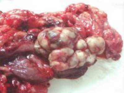 |
Sequence 10 of 11.
A Macroscopic view of a lobulated colon tumor, which
was firm and pseudo encapsulated.
|
|
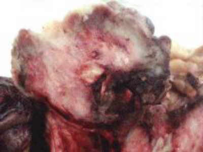 |
Sequence 11 of 11.
Here, the cut surface is shown, redish and limited.
|
|
 |
Video Endoscopic Sequence 1 of 3.
Metastatic colon adenocarcinoma of the stomach
This is the case of a 72 year-old lady, four years previously undergone surgery due to a adenocarcinoma of the descending colon limited to the splenic flexure. Two years later the cancer had abdominal recurrence, underwent a new radical cancer surgery an upper endoscopy displays a large mass in the stomach, the biopsies display that are from the colon.
|
|
 |
Video Endoscopic Sequence 2 of 3.
Video endoscopic image of colonic metastasis to the stomach.
|
|
 |
Video Endoscopic Sequence 3 of 3.
Retroflexed image observing the metastases
|
|
 |
Cervix Carcinoma that Infiltrated the Rectum.
Local tumor spread of cervical cancer is currently considered as radial progressive intra- and extracervical permeation. For radical tumor resection or radiation the inclusion of a wide envelope of tumor-free tissue is demanded. However, this concept may lead to considerable treatment-related morbidity and does not prevent local relapse.
|
|
 |
Cervix Carcinoma that Infiltrated the Rectum.
Invasive cervical cancer, The prognosis is based on the stage, size, and histologic grade of the primary tumor and the status of the lymph nodes. Assessment of the stage of disease is important in determining whether the patientmay benefit from surgery or will receive radiation therapy.
|
|
 |
Protuded Adenocarcinoma of Prostate into the Rectum
Carcinoma of the prostate was suspected because of other clinical features and this was confirmed by biopsy.
|
|
 |
Video Endoscopic Sequence 1 of 3.
Mucinous cystadenocarcinoma of the ovary that infiltrates the rectum.
This the case of a 53-year old female that
5- year previously, underwent a surgery and chemotherapy
due to a mucinous cystadenocarcinoma of the ovary.
|
|
 |
Video Endoscopic Sequence 2 of 3.
Colonoscopy of Mucinous cystadenocarcinoma of the ovary that infiltrates the rectum.
A mucinous cystadenocarcinoma of the ovary is a rare
malignant form of ovarian mucinous tumours. This type can
account for 5 - 10 % of all ovarian mucinous tumours. It is
a type of ovarian epithelial tumour.
Most well-differentiated mucinous ovarian
adenocarcinomas show mostly expansile invasion or a
confluent glandular growth pattern without destructive
stromal invasion. Expansile invasion, first formally
described in mucinous adenocarcinomas, refers to
architecturally complex glandular arrangements
incompatible with a noninvasive process.
|
|
 |
Video Endoscopic Sequence 3 of 3.
Endoscopic Image of Mucinous cystadenocarcinoma of the ovary that infiltrates the rectum.
Pathology
Sub types
Mucinous ovarian tumours can be broadly sub classified into 3 main sub groups
ovarian mucinous cystadenoma : 80 %
ovarian borderline mucinous tumour : 10 - 15 %
ovarian mucinous cystadenocarcinoma : 5 - 10 %
|
|
|
|
|
|
|