 |
|
 |
Video Endoscopic Sequence 1 of 20.
Prolasep Rectal adenocarcinoma resected with endoscopic endoresection
A 52-year-old female who for for the previous ten months had presented with a prolapsed mass.
Despite the fact that colorectal polyps and solitary rectal ulcers may be present in conjunction with rectal prolapse, association between rectal prolapse and rectal cancer is very rare.

All endoscopic images in this Atlas contain a video, which can be downloaded to your slide and used in academic presentations. |
|
 |
Video Endoscopic Sequence 2 of 20.
Another image and video of the mass prolapsed through the anus. |
|
.") |
Video Endoscopic Sequence 3 of 20.
Ten days after her first apoiment, an endoscopic resection was performed. The biopsies following this resection revealed: adenocarcinoma originating from a tubulovillous polyp. With (Multiple foci of adenocarcinoma).
|
|
 |
Video Endoscopic Sequence 4 of 20.
Another image and video clip of the endoresection
|
|
 |
Video Endoscopic Sequence 5 of 20.
The image and video clip were obtained with an extra video camera; three large fragments of the tumor resected with a diathermy loop can be seen.
|
|
 |
Video Endoscopic Sequence 6 of 20.
The second fragment of the tumor is visible.
|
|
 |
Video Endoscopic Sequence 7 of 20.
The third fragment of the tumor is visible.
|
|
 |
Video Endoscopic Sequence 8 of 20.
Retroflexed image
A multilobulated mass is observed; this image and video clip are from before the start of the endoscopic endoresection.
|
|
 |
Video Endoscopic Sequence 9 of 20..
The mass is observed through the anal canal.
|
|
 |
Video Endoscopic Sequence 10 of 20.
Endoscopic endoresection using a diathermy loop
|
|
 |
Video Endoscopic Sequence 11 of 20.
Endoscopic Resection of Rectal Neoplasia.
|
|
 |
Video Endoscopic Sequence 12 of 20.
Endoscopic Resection of Rectal Neoplasia. |
|
 |
Video Endoscopic Sequence 13 of 20.
Twenty days after the first endoscopic resection, a new therapeutic colonoscopy is performed.
The status after endoscopic endoresection is observed; there are some fragments of adenomas and scars. Small fragments are then resected.
|
|
 |
Video Endoscopic Sequence 14 of 20.
Saline solution is infiltrated into the residual fragments
|
|
 |
Video Endoscopic Sequence 15 of 20.
Residual fragments of adenomatous tissue are resected using a diathermy loop.
|
|
 |
Video Endoscopic Sequence 16 of 20.
Continue to dry the remaining fragments with a diathermy loop. |
|
 |
Video Endoscopic Sequence 17 of 20.
More fragments have been resected |
|
 |
Video Endoscopic Sequence 18 of 20.
Argon Plasma Coagulator is used to cauterize any remaining cells. |
|
 |
Video Endoscopic Sequence 19 of 20.
Final status of endoscopic endoresection of a rectal adenocarcinoma |
|
 |
Video Endoscopic Sequence 20 of 20.
Final status of endoscopic endoresection of a rectal adenocarcinoma |
|
 |
Endoscopic polypectomy.
Colonoscopy and endoscopic polypectomy are the most powerful tools for preventing colon carcinoma.
|
|
 |
Video Endoscopic Sequence 1 of 8.
Polypectomy of Pedunculated Rectal Polyp
This 41-year-old male patient was found to have this polyp during a routine colonoscopy.
|
|
 |
Video Endoscopic Sequence 2 of 8.
The polypectomy is performed using a diathermy loop.
|
|
 |
Video Endoscopic Sequence 3 of 8.
I |
|
 |
Video Endoscopic Sequence 4 of 8.
. |
|
 |
Video Endoscopic Sequence 5 of 8.
. |
|
 |
Video Endoscopic Sequence 6 of 8.
|
|
 |
Video Endoscopic Sequence 7 of 8.
|
|
 |
Video Endoscopic Sequence 8 of 8.
Two years later a new colonoscopy was performed, revealing the scar of the polypectomy. |
|
 |
Video Endoscopic Sequence 1 of 23.
A 43-year-old female patient presented with rectal bleeding of approximately three months. A colonoscopy revealed an adenomatous polyp approximately two centimeters in size with a long, wide pedicle.
It was removed via endoscopic polypectomy. |
|
 |
Video Endoscopic Sequence 2 of 23.
Another image of such a mass.
|
|
 |
Video Endoscopic Sequence 3 of 23.
The pedicle is observed to be large and wide
|
|
 |
Video Endoscopic Sequence 4 of 23.
The image and video show it being observed with chromatoscopy using indigo carmine. |
|
 |
Video Endoscopic Sequence 5 of 23.
This image and video show a colonoscope with magnification. |
|
 |
Video Endoscopic Sequence 6 of 23.
Another image using chromatoscopy
|
|
 |
Video Endoscopic Sequence 7 of 23.
Amoebic Ulcer..
|
|
 |
Video Endoscopic Sequence 8 of 23.
Infiltration with adrenaline in saline solution at 1/10.000
|
|
 |
Video Endoscopic Sequence 9 of 23.
Infiltration with adrenaline in saline solution at
1/10.000
|
|
 |
Video Endoscopic Sequence 10 of 23.
After the infiltrates,the stalk turns white.
|
|
 |
Video Endoscopic Sequence 11 of 23.
A triclip is applied to the stalk.
|
|
 |
Video Endoscopic Sequence 12 of 23.
|
|
 |
Video Endoscopic Sequence 13 of 23.
A second triclip is placed
|
|
 |
Video Endoscopic Sequence 14 of 23.
The two triclips are visible on the pedicle
|
|
 |
Video Endoscopic Sequence 15 of 23.
. |
|
 |
Video Endoscopic Sequence 16 of 23.
Endoscopic polypectomy with a diathermy loop
. |
|
 |
Video Endoscopic Sequence 17 of 23.
|
|
 |
Video Endoscopic Sequence 18 of 23..
|
|
 |
Video Endoscopic Sequence 19 of 23.
|
|
 |
Video Endoscopic Sequence 20 of 23.
|
|
 |
Video Endoscopic Sequence 21 of 23.
|
|
 |
Video Endoscopic Sequence 23 of 23. |
|
 |
Video Endoscopic Sequence 23 of 23. |
|
 |
Video Endoscopic Sequence 1 of 6.
|
|
 |
Video Endoscopic Sequence 2 of 6..
|
|
 |
Video Endoscopic Sequence 3 of 6.
|
|
 |
Video Endoscopic Sequence 4 of 6.
|
|
 |
Video Endoscopic Sequence 5 of 6.
|
|
 |
Video Endoscopic Sequence 6 of 6.
|
|
 |
Video Endoscopic Sequence 1 of 4.
|
|
 |
Video Endoscopic Sequence 2 of 4.
|
|
 |
Video Endoscopic Sequence 3 of 4.
. |
|
 |
Video Endoscopic Sequence 4 of 4.
|
|
 |
Video Endoscopic Sequence 1 of 2.
|
|
 |
Video Endoscopic Sequence 2 of 2.
|
|
 |
Polypectomy of a smal pedunculated polyp
|
|
 |
Video Endoscopic Sequence 1 of 2.
Endoscopic polypectomy.
The snare captures the polyp. |
|
 |
Video Endoscopic Sequence 2 of 2.
. |
|
 |
Video Endoscopic Sequence 1 of 5.
|
|
 |
Video Endoscopic Sequence 2 of 5.
|
|
 |
Video Endoscopic Sequence 3 of 5.
|
|
 |
Video Endoscopic Sequence 4 of 5.
|
|
 |
Video Endoscopic Sequence 5 of 5.
Removing the remaining pedicle. |
|
 |
Polypectomy using argon plasma coagulation
Use of argon plasma coagulator in the removal of the tiny polyp. |
|
 |
Video Endoscopic Sequence 1 of 29
Endoscopic polypectomy of a large pedunculated mass.
A 55-year-old female patient was found to have this mass during a routine medical check-up.
Watch this video on YouTube |
|
 |
Video Endoscopic Sequence 2 of 29
A multilobed mass with a long and wide pedicle is seen. |
|
 |
Video Endoscopic Sequence 3 of 29
Another image of the multilobed mass |
|
 |
Video Endoscopic Sequence 4 of 29.
La imagen y el video muestran el pédiculo largo y ancho. |
|
 |
Video Endoscopic Sequence 5 of 29.
Infiltration with adrenaline at 1/10,000 in 50% dextrose.
|
|
 |
Video Endoscopic Sequence 6 of 29.
More applications of adrenaline dilution for vasoconstrictor effects. |
|
 |
Video Endoscopic Sequence 7 of 29.
Note the color changes of the mass due to the vasoconstriction effect. |
|
 |
Video Endoscopic Sequence 8 of 29. |
|
 |
Video Endoscopic Sequence 9 of 29.
Two hemoclips are applied to the pedicle.. |
|
 |
Video Endoscopic Sequence 10 of 29.
Application of the second hemoclip. |
|
 |
Video Endoscopic Sequence 11 of 29.
After hemostatic measures, endoscopic polypectomy with a diathermy loop is initiated.. |
|
 |
Video Endoscopic Sequence 12 of 29.
These sequences show the cutting effect of the pedicle with the diathermy loop. |
|
 |
Video Endoscopic Sequence 13 of 29.
The polyp must be moved to avoid thermal burns to the contralateral intestinal wall. |
|
 |
Video Endoscopic Sequence 14 of 29.
The tumor has already been cut out but it has not completely detached. |
|
 |
Video Endoscopic Sequence 15 of 29.
Again, using diathermy loop, they try to remove it once more. |
|
 |
Video Endoscopic Sequence 16 of 29.
Having detached, the remnant of the pedicle with the two hemoclips can be seen. |
|
 |
Video Endoscopic Sequence 17 of 29.
The mass is observed through remnants of secretions.
|
|
 |
Video Endoscopic Sequence 18 of 29.
|
|
 |
Video Endoscopic Sequence 19 of 29
Photographs of the cut mass.
Multinodulated polypoid tumor measuring 4 x 1.5 x 1.5 cm.. |
|
 |
Video Endoscopic Sequence 20 of 29.
Otra imágenes de la masa. |
|
 |
Video Endoscopic Sequence 21 of 29.
Multilobulated tumor with focal areas of ulceration. |
|
 |
Video Endoscopic Sequence 22 of 29.
More images of the multilobed mass. |
|
 |
Video Endoscopic Sequence 23 of 29.
Más imágenes de la masa multilobulada. |
|
 |
Video Endoscopic Sequence 24 of 29.
Lobed polyp with sessile base. |
|
 |
Video Endoscopic Sequence 25 of 29.
|
|
 |
Video Endoscopic Sequence 26 of 29.
Tubulovellous adenoma of the descending colon.
Normal colonic mucosa is visible at the base of the polyp.
Under the microscope, it is a tubulovillous neoplasm of colonic mucosa with fibrous stroma; there are superficial ulcerated areas; the pedicle has normal muscularis mucosae and colonic mucosa; no atypia or malignancy was found. |
|
 |
Video Endoscopic Sequence 27 of 29.
Tubulovillous pattern at low magnification. |
|
 |
Video Endoscopic Sequence 28 of 29.
A more detailed look at the histological aspect. |
|
 |
Video Endoscopic Sequence 29 of 29.
Tubulovillous pattern with vascular connective tissue axis. |
|
 |
Video Endoscopic Sequence 1 of 35.
Endoscopic Resection of Giant Tubulovillous Adenoma.
This is the case of a 57-year-old lady who had been experiencing rectal bleeding during six months and had only been prescribed ointments and suppositories. She was referred to our unit for evaluation. Anoscopy revealed a mass that initially appeared small, but a rectal examination showed it could be prolapsed through the anal canal. We immediately prepared her with potent laxatives for a colonoscopy and polypectomy. |
|
 |
Video Endoscopic Sequence 2 of 35.
After administering oral laxatives, we decided to examine him and remove the tumor endoscopically
Large polyps are indeed associated with a higher rate of complications. Some polyps require surgical removal; the best indicator of whether a particular endoscopist should attempt removal is their level of experience and how confident they are in their skill and expertise.
One should not assume that because one's own comfort level is exceeded, this surgery is necessarily a mandate, in which case, the endoscopist should be prepared to consult with a more experienced colleague or consider referring the patient to another center. |
|
 |
Video Endoscopic Sequence 3 of 35.
Upon performing retroflexion in the rectum, a large mass is observed that appears to be adenocarcinoma.
At this time we had no pathology results since we decided to remove this neoplasm which we initially thought was small but had a soft appearance. |
|
 |
Video Endoscopic Sequence 4 of 35.
Another image of the irregularly shaped mass
Which patients should initially be treated with surgical resection? |
|
 |
Video Endoscopic Sequence 5 of 35.
Since the initial plan was to remove the tumor, which appeared small, we dared to perform a fragmented polypectomy because we did not have any biopsy reported by pathology. |
|
 |
Video Endoscopic Sequence 6 of 35.
When looping the diathermy loop, the large fragment prolapses to the outside and we perform polypectomy of this fragment outside the anal margin. |
|
 |
Video Endoscopic Sequence 7 of 35.
Multiple fragments of the neoplasm with a hairy appearance are observed.
Note: In this endoscopic resection of this tumor, we did not use any injected fluid to lift the mucosa because the rectal walls are quite thick and there is no danger of perforation as could happen in the rest of the colon. |
|
 |
Video Endoscopic Sequence 8 of 35.
Another image and video observing the infiltrative-looking fragments. |
|
 |
Video Endoscopic Sequence 9 of 35.
Then we removed another large fragment |
|
 |
Video Endoscopic Sequence 10 of 35.
Active bleeding is observed |
|
 |
Video Endoscopic Sequence 11 of 35.
To stop this bleeding using the argon plasma, stopping the bleeding. |
|
 |
Video Endoscopic Sequence 12 of 35.
Continuing to resect the neoplasm by removing several remnants with the diathermy loop. |
|
 |
Video Endoscopic Sequence 13 of 35.
More fragments are being resected with the diathermy loop |
|
 |
Video Endoscopic Sequence 14 of 35.
Some fragments are attached to the pectinate line; we continue using the diathermy loop to cut these fragments with cautery. In this anatomical region, care must be taken when performing endoscopic polypectomy due to the pain that cautery can cause in the anal tissue. |
|
 |
Video Endoscopic Sequence 15 of 35.
Fragments of this giant tubulo villous adenoma continue to be resected in the pectineal line. |
|
 |
Video Endoscopic Sequence 16 of 35. |
|
 |
Video Endoscopic Sequence 17 of 35.
Continuing with the polypectomy of large fragments, retroflexion maneuver. |
|
 |
Video Endoscopic Sequence 18 of 35.
his image and video clip show that the tumor has almost completely Excised. |
|
 |
Video Endoscopic Sequence 19 of 35.
TAnother large fragment. |
|
 |
Video Endoscopic Sequence 20 of 35.
The fragment is placed in the formalin container. |
|
 |
Video Endoscopic Sequence 21 of 35.
View after endoscopic resection of the tumor
This image and video clip show the status of the endoscopic resection of this giant tubulovillous tumor of the rectum. |
|
 |
Video Endoscopic Sequence 22 of 35.
Cauterization is applied with argon plasma coagulator of micro remnants and hemostasis is performed. |
|
 |
Video Endoscopic Sequence 23 of 35.
Final status of endoscopic resection.
Two weeks later, a new endoscopy is performed, see below. |
|
 |
Video Endoscopic Sequence 24 of 35.
Photograph of some of the fragments
Macroscopic details of the excised tumor show a hairy appearance with a dark red surface. |
|
 |
Video Endoscopic Sequence 25 of 35.
Base of the polyp with vascular connective tissue and thermal effects
|
|
 |
Video Endoscopic Sequence 26 of 35.
Panoramic view of the tubulovillous pattern. |
|
 |
Video Endoscopic Sequence 27 of 35.
Detail with greater increase in hair size. |
|
 |
Video Endoscopic Sequence 28 of 35.
High magnification detail of cellular atypia. |
|
 |
Video Endoscopic Sequence 29 of 35.
The polyp mucosa and submucosa with the muscularis mucosae can be seen.
|
|
 |
Video Endoscopic Sequence 30 of 35.
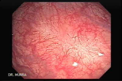
Follow-up endoscopy two weeks after endoscopic resection of the tumor, tissue in the healing phase is observed, multiple biopsies were taken demonstrating only granulation tissue. |
|
 |
Video Endoscopic Sequence 31 of 35..
Final stage of healing. |
|
 |
Video Endoscopic Sequence 32 of 35.
Front view of the scar. |
|
 |
Video Endoscopic Sequence 33 of 35.
Reinforcing with the argon plasma coagulation therapy in case there are any remnants of adenomatous tissue. |
|
 |
Video Endoscopic Sequence 34 of 35
Continuing with ablative therapy. |
|
 |
Video Endoscopic Sequence 35 of 35.
. |
|
|
|
|
|
|
