 |
|
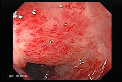 |
Video Endoscopic Sequence 1 of 13.
Post-Radiation Colitis.
This 77 year old woman underwent radiation therapy for cervical cancer approximately one and a half years ago. She remained well over one year. But over the past five months, she has had increasingly frequent episodes of rectal bleeding requiring two monthly blood transfusions until she was referred to our clinic for specific treatment. Colonoscopy revealed fine, tortuous blood vessels and telangiectasias in the rectum.
Download the video clips by clicking on the endoscopic images, if you wish to observe in full screen, wait to bedownloaded complete then press Alt and Enter for Windows media, Real Player Ctrl and 3. Configure the windows media in repeat is optimal. All endoscopic images shown in this Atlas contain video clips. We recommend seeing the video clips in full screen mode.
|
|
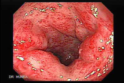 |
Video Endoscopic Sequence 2 of 13.
Endoscopic view of Post-irradiation proctitis
Post-irradiation proctitis with extensive rectal musocal hypervascularity.
The image and the video clip show evidence of radiation injury. The changes seen here are classic for radiation induced proctitis.
|
|
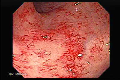 |
Video Endoscopic Sequence 3 of 13.
(Post-irradiation mucosal vascular changes).
Colonoscopy demonstrates multiple twisted small telangiectasias compatible with her history of radiation exposure.
The most common symptom is rectal bleeding inassociation with rectal inflammation known as radiation proctitis. The bleeding is caused by new blood vessel formation as a result of the inflammation caused by radiation. These blood vessels are fragile and bleed easily.
|
|
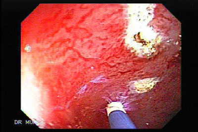 |
Video Endoscopic Sequence 4 of 13.
The patient was given therapy with argon plasma coagulation therapy.
Radiation colitis, an insidious, progressive disease of increasing frequency, develops 6 mo to 5 years after regional radiotherapy for malignancy, owing to the deleterious effects of the latter on the colon and the small intestine. When dealing with radiation colitis and its complications, the most conservative modality should be employed because the areas of intestinal injury do not tend to heal. Acute radiation colitis is mostly self-limited, and usually, only supportive management is required. Chronic radiation colitis, a poorly predictable progressive disease, is considered as a precancerous lesion; radiation-associated malignancy has a tendency to be diagnosed at an advanced stage and to bear a dismal prognosis. Therefore, management of chronic radiation colitis remains a major challenge owing to the progressive evolution of the disease, including development of fibrosis, endarteritis, edema, fragility, perforation, partial obstruction, and cancer. Patients are commonly managed conservatively. Surgical intervention is difficult to perform because of the extension of fibrosis and alterations in the gut and mesentery, and should be reserved for intestinal obstruction, perforation, fistulas, and severe bleeding. Owing to the difficulty in managing the complications of acute and chronic radiation colitis, particular attention should be focused onto the prevention strategies. Uncovering the fibrosis mechanisms and the molecular events underlying radiation bowel disease could lead to the introduction of new therapeutic and/or preventive approaches. A variety of novel, mostly experimental, agents have been used mainly as a prophylaxis, and improvements have been made in radiotherapy delivery, including techniques to reduce the amount of exposed intestine in the radiation field, as a critical strategy for prevention. |
|
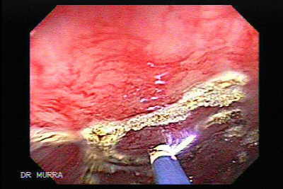 |
Video Endoscopic Sequence 5 of 13.
Non-contact method of coagulation using the APC probe. APC is a non-contact thermal device.
A high voltage spark is delivered at the tip of the probe, which ionizes the argon gas as it is sprayed from the probe tip in the direction of the target tissue. Argon gas is non-flammable and inexpensive to refill. It is easily ionized by the 6000 volt peak energy delivered by the tungsten wire that terminates just proximal to the probe tip. This ionized gas or plasma then seeks a ground in the nearest tissue, delivering the thermal energy with a depth of penetration of roughly 2 to 3 mm. The plasma coagulates both linearly and tangentially. By delivering energy to all tissue near the probe tip which will conduct electricity, APC can be used to treat a lesion around a fold and not clearly in view or en face.
|
|
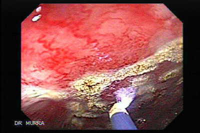 |
Video Endoscopic Sequence 6 of 13.
Argon Plasma Coagulation is an Effective Treatment for Refractory Hemorrhagic Radiation Proctitis.
|
|
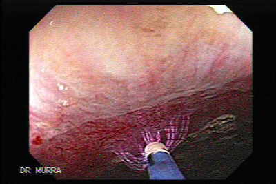 |
Video Endoscopic Sequence 7 of 13.
One of the most effective treatments for radiation proctitis is Argon Plasma Coagulation.
|
|
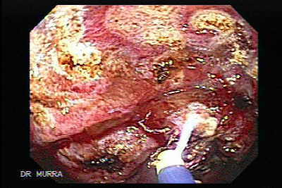 |
Video Endoscopic Sequence 8 of 13.
A forward firing probe is used to fulgurate individual sites.
Firstly, argon gas is emitted from the end of the probe running through the endoscope channel. Next, high-frequency current is discharged from an electrosurgery unit. When argon gas becomes electrically conductive ( argon plasma ) this allows the current to reach the targeted mucosa of the tissue which is coagulated shallowly and uniformly.
The device is especially effective for the coagulation of hemorrhages on the surface of the tissue.
|
|
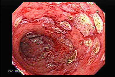 |
Video Endoscopic Sequence 9 of 13.
This image as well as the video clips display multiple post treatment ulcers.
The goal of endoscopic therapy is to ablate the angioectasias with a resultant improvement in the severity and frequency of rectal bleeding. This will lead to an increase in the hemoglobin level, reduction in transfusion requirements and hopefully, an improvement in quality of life. Candidates for endoscopic therapy include, but are not limited to, patients with chronic hematochezia associated with transfusion dependency.
|
|
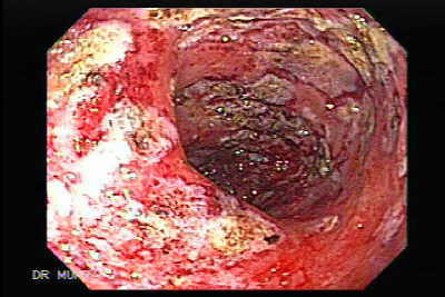 |
Video Endoscopic Sequence 10 of 13.
The procedures described above are considered to be safe. However, temporary discomfort or pain may occur following introduction of air into the stomach or bowel. Major complications are rare but can occur. These complications include perforation.
|
|
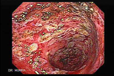 |
Video Endoscopic Sequence 11 of 13.
Therapy with argon plasma coagulator.
It conducts monopolar electrosurgical current to tissue via an ionized argon gas stream (argon plasma) that is delivered from a small tube that emanates from the colonoscope. The argon gas is ignited and charged and cause a superficial destruction of these abnormal blood vessels and stops the radiation related bleeding. The APC procedure is performed on an outpatient basis and requires only light or no sedation. APC is consider not only effective, but very safe.
|
|
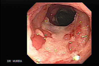 |
Video Endoscopic Sequence 12 of 13.
APC sessions can be repeated as needed to control bleeding.
Nine weeks later, the ulcers have healed and only a few Lesions remain. Her bleeding has almost completely ceased. She does not need to be seen in follow up unless bleeding recurs.
|
|
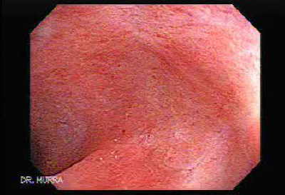 |
Video Endoscopic Sequence 13 of 13.
Six months later, the nearly complete resolution of the process is observed.
|
|
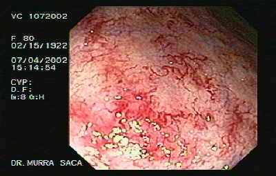 |
Video Endoscopic Sequence 1 of 4.
Post-Radiation Colitis.
An 80 year-old woman with post-radiation proctitis. The patient has cervix carcinoma and underwent radiotherapy. The images and videos display telangiectasias, which is the physiopathologic cause of this clinical entity.
In 1897, 2 years after the discovery of x-rays by Roentgen, radiation-induced intestinal injury was first reported.
|
|
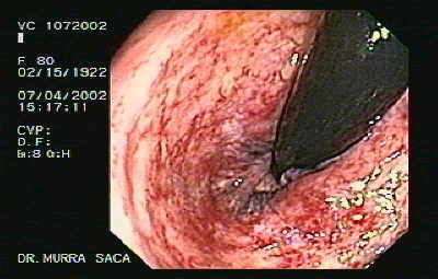 |
Video Endoscopic Sequence 2 of 4.
Hemorrhagic radiation proctitis. Retroflexed image
The proctitis has affected only 3 centimeters above the pectinea line.
Radiation injury is the damage to the soft tissue and bone,following radiation therapy. There are early and late effects (that occur weeks and years after the radiation) caused by ischemia, interruption of the blood supply and direct cells damage, which are sensitive to radiation.
|
|
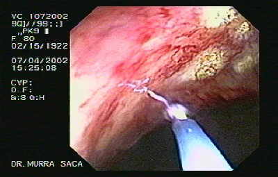 |
Video Endoscopic Sequence 3 of 4.
Therapy with argon plasma coagulator (APC).
Argon Plasma Coagulation, or APC for short, is a new method of electrocoagulation. As a result, it allows for the non-contact application of electrical energy to achieve tissue destruction or hemostasis (the ability to stop bleeding). APC uses high frequency electrical current delivered via ionized argon gas. This gas, being ionized, allows for the conduction of electricity, thus leading to the term "argon plasma"
|
|
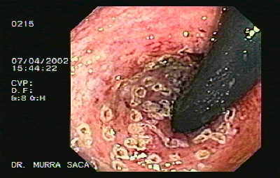 |
Video Endoscopic Sequence 4 of 4.
Status post the argon treatment, image in retrofletion.
Radiation has been used in the treatment of cancers since
the 1930's. It still remains the mainstay of treatment for
cervical and vaginal carcinomas around the world. Its role
in the treatment of other gynaecological cancers is less
assured. It will probably continue to play a role in the
management of some endometrial and vulvar carcinomas.
Radiotherapy may be the only treatment or part of a
multidisciplinary treatment plan, this reflects the modern
approach to the management of gynaecological
cancers.
|
|
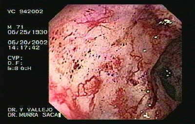 |
Video Endoscopic Sequence 1 of 14.
Radiation Proctosigmoiditis.
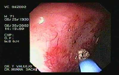
Post-irradiation proctopathy in a 71 year-old man, who underwent radiation therapy for prostatic carcinoma. The image and the video display hemorrhage, clots and telangiectasias.
|
|
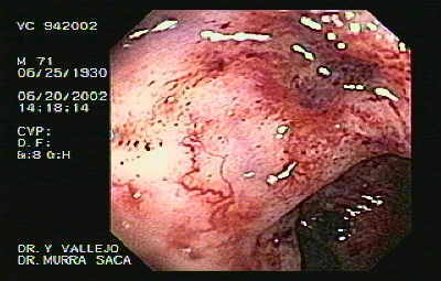 |
Video Endoscopic Sequence 2 of 14.
Radiation Proctosigmoiditis.
The typical endoscopic appearance is characterized by
telangiectasias and hemorrhagic mucosal changes.
The symptoms may arise months to years after the end of
radiotherapy and mainly include tenesmus with discharge
of mucus and blood.
|
|
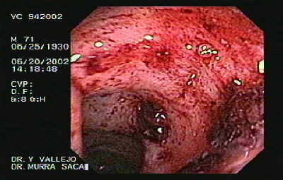 |
Video Endoscopic Sequence 3 of 14.
Radiation Proctitis.
Post-irradiation proctopathy in a 71 year-old man, who had underwent radiation therapy for prostatic carcinoma.
The image and the video display hemorrhage, clots and telangiectasias.
|
|
 |
Video Endoscopic Sequence 4 of 14.
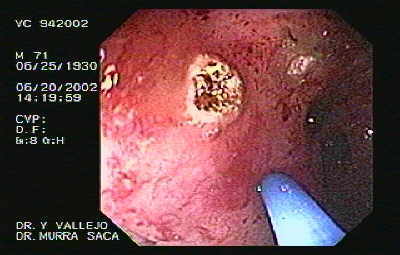
The patient underwent radiotherapy. Six months later that patient started with rectal discharge.
Argon Plasma Coagulation beam, in a case of post radiation proctitis, due to prostatic carcinoma 3 years before. |
|
 |
Video Endoscopic Sequence 5 of 14.
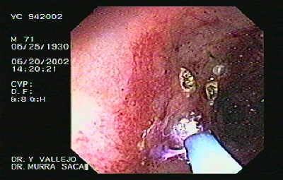
Argon Plasma Coagulation treatment, in a case of post radiation proctitis, due to prostatic carcinoma. Radiation proctosigmoiditis is a frequent complication of radiation therapy, used in the treatment of cervix or uterus cancer in women, prostate and testes cancer in men and urine bladder in both sexes. Eradication of hypervascularity using an argon plasma coagulator. A single pulse has been delivered, with a visiblecoagulation effect.
|
|
 |
Video Endoscopic Sequence 6 of 14.
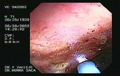
Radiation is a common therapy administered to patients with malignant disease in the pelvis, within or outside the abdomen. It may be administered alone or in conjunction with various types of chemotherapy, some of which increase the risk of radiation injury to the gastrointestinal tract.
|
|
 |
Video Endoscopic Sequence 7 of 14.
As many as half of the patients with various cancers receive some form of radiation. One to three weeks later, almost 50 to 75 percent experienced a subacute gastrointestinal syndrome. An estimated 0.5 to 17 percent (average: 5 percent) go on to develop chronic radiation enterocolitis from nine months later up to 20 years after radiation therapy.
|
|
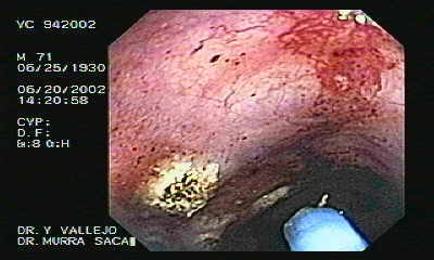 |
Video Endoscopic Sequence 8 of 14.
Radiation Proctitis.
|
|
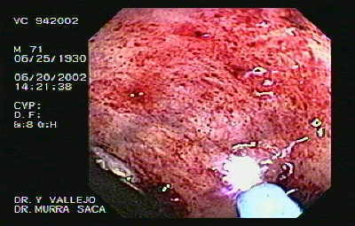 |
Video Endoscopic Sequence 9 of 14.
The image and the video display the argon plasma coagulator beam inside of the rectal tissue. The efficacy and safety of argon plasma coagulation therapy in the post radiation colitis is becoming as the best therapy available for radiotherapy´s “secondary effects”.
|
|
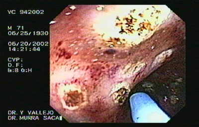 |
Video Endoscopic Sequence 10 of 14.
Multiple applications of argon beam are observed.
|
|
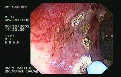 |
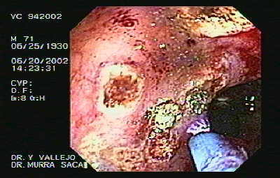
Video Endoscopic Sequence 11 of 14.
Sequelae of Radiotherapy.
The incidence of severe complications rises with the total dose. The higher the dose the better the local control of the disease; however the complication rate is also higher. Bowel damage ranges from diarrhea and rectal irritation to perforation, obstruction and severe rectal ulceration. Bladder damage may result in a vesico-vaginal fistula.
|
|
 |
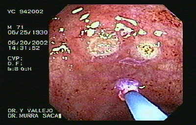
Video Endoscopic Sequence 12 of 14.
Various factors are associated with a high risk of late radiation complications: advanced age, hypertension, anaemia, poor nutrition status, diabetes mellitus, obesity, previous abdominal or pelvic surgery, pelvic inflammatory disease, colitis and diverticulitis. If any of these factors are present, the total dose may need to be reduced to minimize the likelihood of complications.
|
|
 |
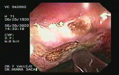
Video Endoscopic Sequence 13 of 14.
The effectiveness of argon coagulator for radiation proctitis has been established in many clinical trials.
|
|
 |
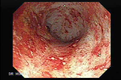
Video Endoscopic Sequence 14 of 14.
Lesions are observed during water lavage.
Radiation-induced injury is best described in 2 ways. Acute injury is a function of fractionation of the dose, field size, type of radiation, and frequency of treatment. Acute injury is caused by injury to the mitotically active intestinal crypt cells. On the other hand, chronic radiation injury is caused by injury to the less mitotically active vascular endothelial and connective tissue cells. Chronic injury is a function of the total dose of radiation used. This accounts for the described biphasic radiation injury.
|
|
 |
Video Endoscopic Sequence 1 of 4.
Radiation Enteritis
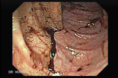
This 74 year-old, female, underwent radiation therapy for uterine cervix cancer 6 month after iniciates with rectal bleeding, requiring red blood cell transfusion, a colonoscopy was performed finding the image here presented.
|
|
 |
Video Endoscopic Sequence 2 of 4.
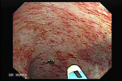
In the sigmoid there are some rest of blood that seem to be melena, radiation enteritis was detected in the jejuno the first lesion was found at 150 cm from the Triez angle and the second 200 cm. In spite of argon plasma coagulation therapy, patient continues with bleeding with melena, and multiple transfusions of blood was performed, the upper endoscopy was negative and a angiotac was done finding two areas of bleeding in the jejuno, patient underwent surgery of the jejuno, a 30 cm bowel resection was performed due to the severe ischemic damage of jejuno with post irradiation enteritis, (unusual case of radiation enteritis).
|
|
 |
Video Endoscopic Sequence 3 of 4.
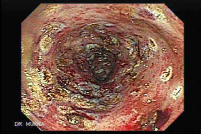
Multiple applications of argon beam are observed.
Radiation therapy is a powerful method for the control of cancer. The utilization of abdominal or pelvic radiation has been extended, and the incidence of radiation enteritis appears to be increasing. The majority of the induced lesions is in the distal ileum, sigmoid colon, or rectum.
|
|
 |
Video Endoscopic Sequence 4 of 4.
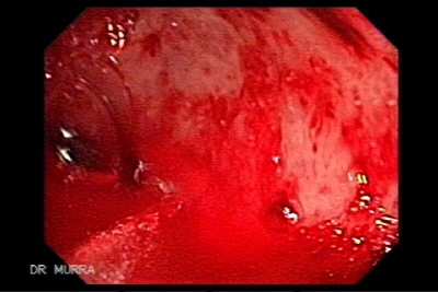
Status post the argon plasma therapy
|
|
 |
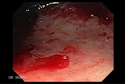
Video Endoscopic Sequence 1 of 12.
Radiation Colitis with active micro-hemorrhages
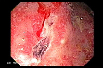
This a 72 year-old male with alcoholic liver cirrhosis. Who had received radiotherapy with cobalt for prostate cancer. After four previous months, begins with rectal bleeding, had received blood transfusion at another clinic, the patient was referred to our endoscopic unit for appropriate treatment.
The interesting thing about this case is that colonoscopy are finding several micro active bleeding in the rectal area near the dentate line.
Due to acute bleeding the first colonoscopy, It was prepared with local enemas, For the next day the colon was prepared with oral laxatives for the second colonoscopy. |
|
 |
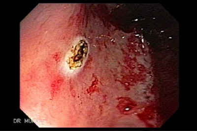
Video Endoscopic Sequence 2 of 12.
Radiation Colitis with active micro-hemorrhages
It is recommended to analyze the video clip, because although we have had many cases of post radiation colitis. We had not previously encountered similar cases with micro active bleeding. Perhaps coagulation it may have some role the due to a his liver disease.
|
|
 |
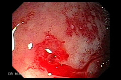
Video Endoscopic Sequence 3 of 12.
Radiation Colitis with active micro-hemorrhages
It is cauterized with monopolar cautery, For the others bleeding, the argon plasma coagulation is used.
|
|
 |
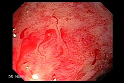
Video Endoscopic Sequence 4 of 12.
It is observed more active micro-hemorrhages |
|
 |
Video Endoscopic Sequence 5 of 12.
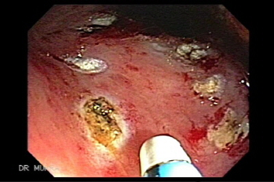
In this image as well as the video clip the are more and active micro-hemorrhages
|
|
 |
Video Endoscopic Sequence 6 of 12.
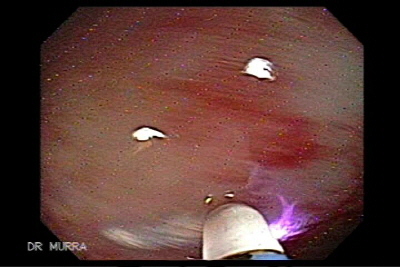
Radiation colitis is a common consequence of pelvic radiation. Its complications may include anemia due to chronic bleeding requiring transfusions. Many of these patients are managed with rectal medications which are often inadequate for control. Argon plasma coagulation (APC) has been well described for its efficacy in treating radiation proctitis. |
|
 |
Video Endoscopic Sequence 7 de 12.
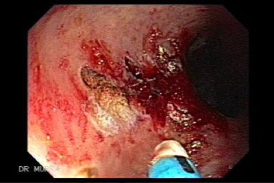
Radiation colitis is a well-recognized complication of radiation therapy for pelvic malignancies, which include prostate, endometrial, cervical or bladder cancers. There are two forms of radiation colitis, acute and chronic. The acute form is usually self-limiting and lasts up to 3 months. The chronic or delayed form may present months or years after radiation exposure. Late complications of pelvic radiotherapy consists of bleeding, anemia, strictures, fistula or anorectal dysfunction, and have an incidence rate of 5–20%. Radiation-induced mucosal damage results in epithelial changes such as fibrosis, microvascular alterations, thrombosis, telangiectasia and endothelial degeneration. Patients often present with persistent rectal bleeding which may lead to iron deficiency anemia requiring blood transfusions. Additionally, reduced distensibility of the rectum due to fibrosis may result in tenesmus and in some cases rectal stricturing leading to difficulty in defecation. |
|
 |
Video Endoscopic Sequence 8 of 12.
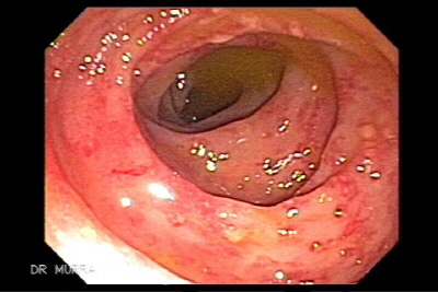
There have been numerous studies on the efficacy of argon plasma coagulation (APC) in the treatment of radiation proctitis. In contrast to conventional unipolar and bipolar electrocoagulation techniques, where adhesion of the probe to tissue and difficulty of assessing depth of thermal effect are major limitations, APC involves the delivery of unipolar diathermy current in a non-contact fashion, using the inert argon gas as the conducting medium. In a study by Sebastian et al. of 25 patients with persistent rectal bleeding and recurrent anemia, 81% achieved complete cessation of bleeding after a median of ne treatment with APC; no complications resulted from the use of this modality. Other studies showed similar efficacies with potential complications such as rectal stricture and recto-vaginal fistula. There have also been case reports of methane gas fire and colonic explosion when using APC. However, complete bowel preparation and a full colonoscopy prior to APC treatment will drastically reduce this risk.
|
|
 |
Video Endoscopic Sequence 9 of 12.
A follow up therapeutic colonoscopy was performed
|
|
 |
Video Endoscopic Sequence 10 of 12.
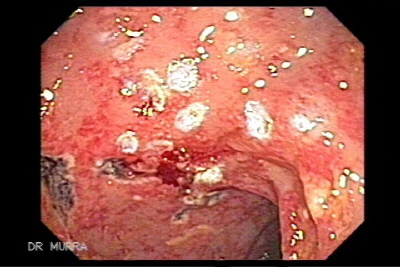
Post-ulcer treatment is observed, the wall has little bleeding which cauterization with APC is applied.
|
|
 |
Video Endoscopic Sequence 11 of 12.
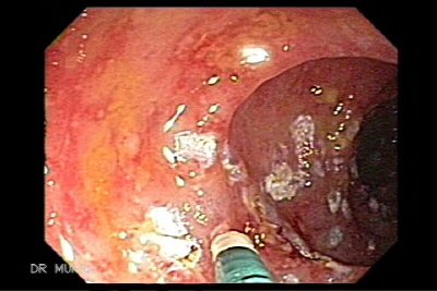
Therapy with argon plasma coagulator.
|
|
 |
Video Endoscopic Sequence 12 of 12.
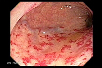
The bleeding stopped but in the near future, due to the extent of lesions of the rectum-sigmoid, He will need more therapy with argon plasma coagulator.
Single sessions have been reported to significantly improve symptoms, but on average, two to three treatments are needed to achieve this result. Improvements persisted for a number of months after therapy was finished.
|
|
 |
Video Endoscopic Sequence 1 of 5
Post-Radiation Colitis.
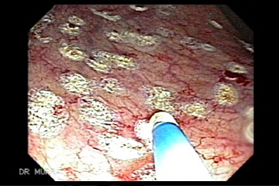
This 71 year old woman underwent radiation therapy for cervical cancer approximately one years ago. She remained well over one year. But over the past five months, she has had increasingly frequent episodes of rectal bleeding requiring two monthly blood transfusions until she was referred to our clinic for specific treatment. Colonoscopy revealed fine, tortuous blood vessels and telangiectasias in the rectum.
|
|
 |
Video Endoscopic Sequence 2 of 5.
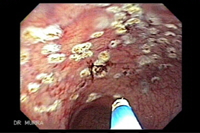
Endoscopic view of Post-irradiation proctitis
Post-irradiation proctitis with extensive rectal musocal hypervascularity.
The image and the video clip show evidence of radiation injury. The changes seen here are classic for radiation induced proctitis.
|
|
 |
Video Endoscopic Sequence 3 of 5.
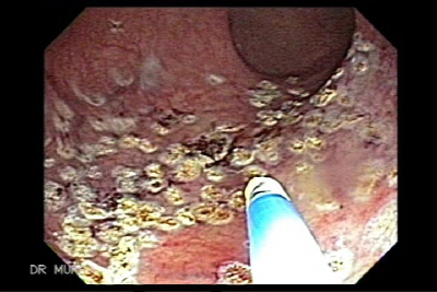
(Post-irradiation mucosal vascular changes).
Colonoscopy demonstrates multiple twisted small telangiectasias compatible with her history of radiation exposure.
The most common symptom is rectal bleeding inassociation with rectal inflammation known as radiation proctitis. The bleeding is caused by new blood vessel formation as a result of the inflammation caused by radiation. These blood vessels are fragile and bleed easily.
|
|
 |
Video Endoscopic Sequence 4 of 5.
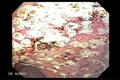
The patient was given therapy with argon plasma coagulation therapy.
|
|
 |
Video Endoscopic Sequence 5 of 5.
Non-contact method of coagulation using the APC probe. APC is a non-contact thermal device.
A high voltage spark is delivered at the tip of the probe, which ionizes the argon gas as it is sprayed from the probe tip in the direction of the target tissue. Argon gas is non-flammable and inexpensive to refill. It is easily ionized by the 6000 volt peak energy delivered by the tungsten wire that terminates just proximal to the probe tip. This ionized gas or plasma then seeks a ground in the nearest tissue, delivering the thermal energy with a depth of penetration of roughly 2 to 3 mm. The plasma coagulates both linearly and tangentially. By delivering energy to all tissue near the probe tip which will conduct electricity, APC can be used to treat a lesion around a fold and not clearly in view or en face.
|
|
 |
Video Endoscopic Sequence 1 of 12.
Post-Radiation Colitis
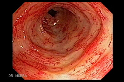
A 69-year-old male patient who had received 33 sessions of radiotherapy with a linear accelerator for prostate cancer.
|
|
| |
Video Endoscopic Sequence 2 of 12.
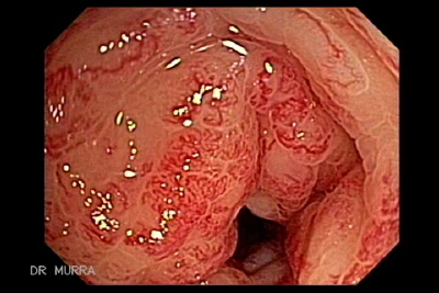
Post Radiation Proctitis with extensive hypervascularity of the rectal mucosa.
The image and the video clip show evidence of radiation injury. The changes visible here are classics of radiation-induced proctitis. |
|
 |
Secuencia Video Endoscópica 3 de 12.
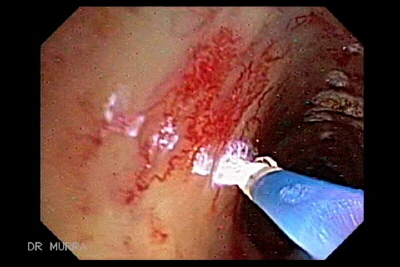
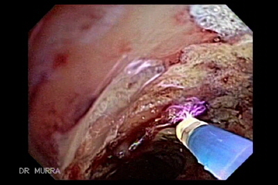
The first session of ablative therapy with argon plasma coagulator (APC) begins.
|
|
 |
Secuencia Video Endoscópica 4 de 12.
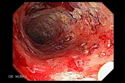
Therapeutic treatment with Argon Plasma Coagulator
|
|
 |
Secuencia Video Endoscópica 5 de 12.
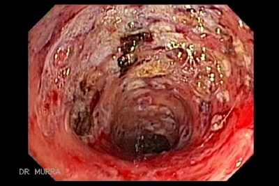
A high voltage spark is delivered at the tip of the probe, which ionizes the argon gas as it is sprayed from the probe tip in the direction of the target tissue.
|
|
 |
Secuencia video endoscópica 6 de 12.
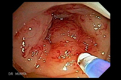
Radiation therapy is a powerful method for the control of cancer. The utilization of abdominal or pelvic radiation has been extended, and the incidence of radiation enteritis appears to be increasing. The majority of the induced lesions is in the distal ileum, sigmoid colon, or rectum. |
|
 |
Video Endoscopic Sequence 7 de 12.
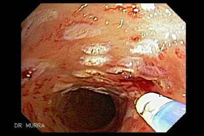
Post irradiation proctitis
Second session of ablative therapy with argon plasma coagulator (APC).
Because the bleeding had decreased but with recurrence, two months later the second session of argon plasma coagulator therapy was performed.
|
|
 |
Secuencia video endoscópica 8 de 12.
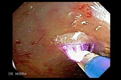
Second session of ablative therapy with argon plasma coagulator (APC). |
|
 |
Secuencia video endoscópica 9 de 12.
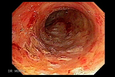
The purple light of the argon plasma coagulator is displayed. |
|
 |
Secuencia video endoscópica 10 de 12.
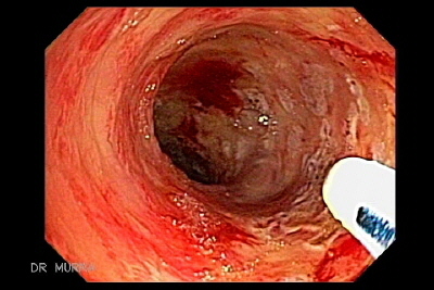
Second session of ablative therapy with argon plasma coagulator (APC).
|
|
 |
Secuencia video endoscópica 11 de 12.
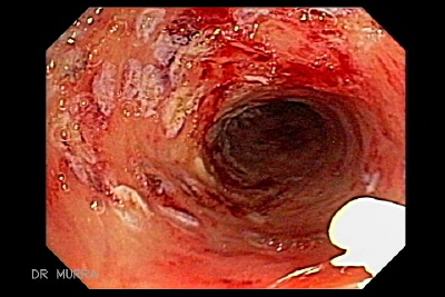
More images and video clips of this case. |
|
 |
Secuencia Video Endoscópica 12 de 12.
Second session of ablative therapy with argon plasma coagulator (APC).
|
|
 |
Secuencia Video Endoscópica 1 de 3.
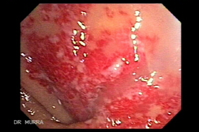
Post irradiation proctitis
A 50-year-old female, who had received 24 sessions of radiotherapy with cobalt for cervical-uterine cancer. Six months later presents recurrent enterorrhagia.
|
|
 |
Secuencia Video Endoscópica 2 de 3.
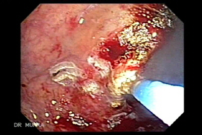
Post irradiation proctitis
A session of ablative therapy with argon plasma coagulator (APC) is initiated.
|
|
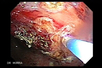 |
Secuencia Video Endoscópica 3 de 3.
Ablative therapy with argon plasma coagulator (APC).
|
|
|
|
|
|
|
